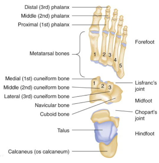
anatomy of left foot
Radiographic Anatomy of the Skeleton: Cervical Spine -- Left Anterior. 16 Pics about Radiographic Anatomy of the Skeleton: Cervical Spine -- Left Anterior : MediVisuals Plantar Anatomy of Left Foot Medical Illustration, How A Simple Foot Adjustment Can Help Your Brain and also MediVisuals Plantar Anatomy of Left Foot Medical Illustration.
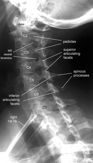
Radiographic Anatomy Of The Skeleton: Cervical Spine -- Left Anterior
 uwmsk.org
spine cervical ray oblique labelled anatomy radiology left radiographic anterior neck diagram skeleton normal imaging vertebrae uwmsk xray schools lateral
MediVisuals Plantar Anatomy Of Left Foot Medical Illustration
 medivisuals1.com
plantar foot anatomy left 01b illustration medivisuals1
Ankle Ultrasound
 fpnotebook.com
ultrasound ankle anterior dorsal technique
Anatomy Of The Lateral Plantar Nerve - Everything You Need To Know - Dr
 www.youtube.com
nerve plantar lateral anatomy
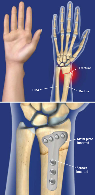
Distal Radius Fracture Repair With Volar Plate | Central Coast
 centralcoastortho.com
radius distal volar fracture plate wrist screws broken fractures repair plates left displaced surgery bones forearm hand portion preparation near
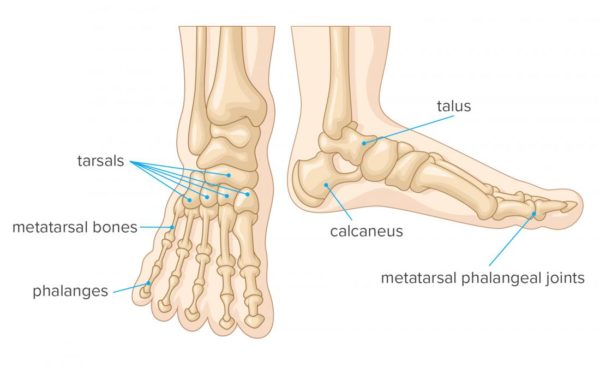
How A Simple Foot Adjustment Can Help Your Brain
 www.yourbodyofknowledge.com
anatomy foot medical illustration feet medicinenet bones joints anatomical structure structures anatomic ankle left right drawing brain simple muscles metatarsal
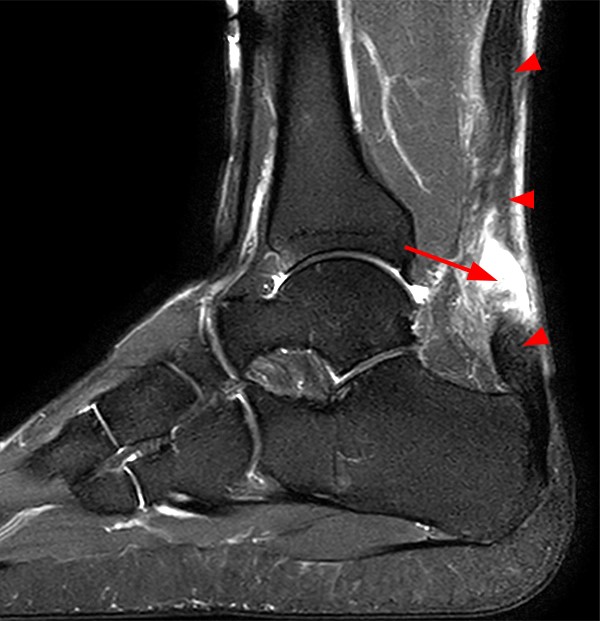
Achilles Tendon Pathology - Radsource
 radsource.us
achilles tendon pathology mri
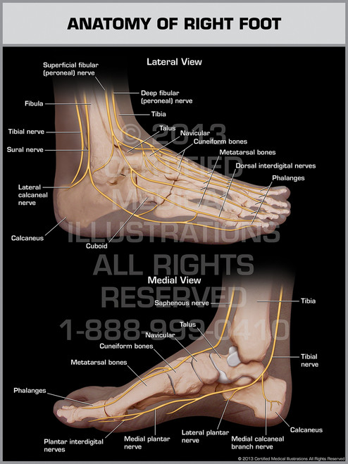
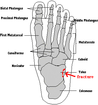
Anatomy Of Right Foot
 certifiedmedicalillustrations.com
right anatomy foot hand forearm leg enlarge
EmDOCs.net – Emergency Medicine EducationFoot Injuries In The Emergency
 www.emdocs.net
foot anatomy 3rd et adults ed emergency emdocs injuries department rockwood york
Foot Pain - Symptoms And Treatment | Home Physio Group
 www.homephysio.com
bones joints spur anatomical medicalnewstoday tarsals phalanges toes metatarsals tulang fungsi jari ligaments symptoms apexbikes fibula tibia tendons
Foot Anatomy
 www.fpnotebook.com
foot anatomy feet hands dig drawing fpnotebook july
Left Foot, Superior View
 www.purposegames.com
foot left superior
Normal Ankle And Foot Anatomy
 www.foot-pain-explained.com
Radiographic Anatomy Of The Skeleton: Lumbar Spine -- Lateral View
 uwmsk.org
spine lumbar lateral radiology ray anatomy labelled diagram coccyx xray radiographic skeleton bones uwmsk human muscle nursing schools medical student
Trip Report: Snap, Crackle, Pop!
 www.swarpa.net
foot anatomy sesamoid bones bone 1st toes under metatarsal snap claw crackle pop physiotherapy oh
Osseous Injuries Of The Foot: An Imaging Review. Part 1: The Forefoot
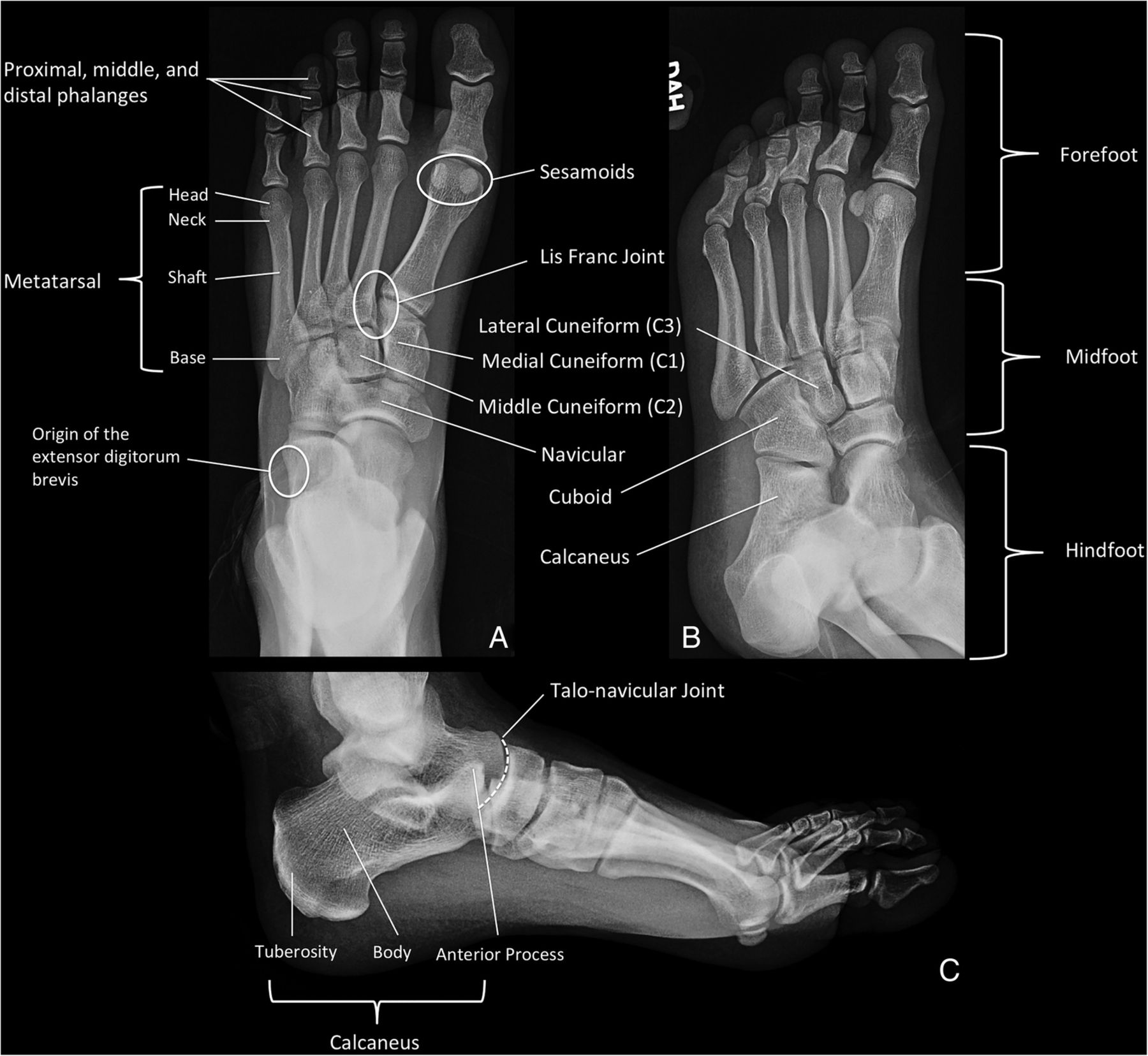 emj.bmj.com
forefoot oblique bmj emj emermed osseous distal
Osseous injuries of the foot: an imaging review. part 1: the forefoot. Forefoot oblique bmj emj emermed osseous distal. Ankle ultrasound