anatomy of left knee
Radiographic Anatomy of the Skeleton: Cervical Spine -- Left Anterior. 11 Pics about Radiographic Anatomy of the Skeleton: Cervical Spine -- Left Anterior : Anatomy Of The Left Knee Diagram, (Hamstrings) Biceps Femoris: Short Head | Chandler Physical Therapy and also CRACKCast E057 – Knee and Lower Leg - CanadiEM.
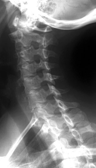
Radiographic Anatomy Of The Skeleton: Cervical Spine -- Left Anterior
 uwmsk.org
spine cervical ray oblique labelled radiology anatomy left radiographic anterior neck diagram skeleton normal imaging vertebrae uwmsk tech xray schools
(Hamstrings) Biceps Femoris: Short Head | Chandler Physical Therapy
 chandlerphysicaltherapy.net
knee patella pain chondromalacia cap anatomy femoris biceps head patellar muscle labeled treatment tendonitis short quadriceps joint femur chiropractor tibia
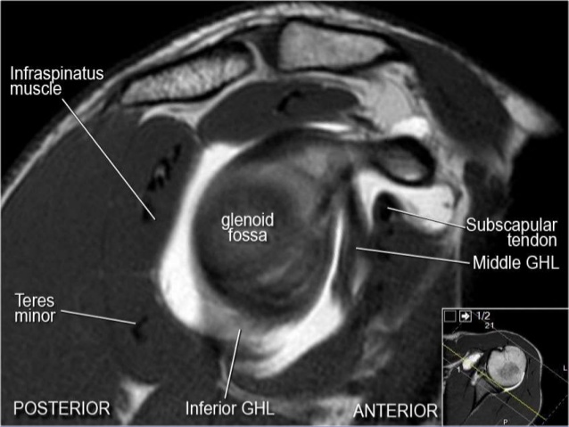
MRI Musculo-Skeletal Section: MRI Anatomy Of The Shoulder (sagittal View).
 mrimusculoskeletalsection.blogspot.com
anatomy mri shoulder sagittal glenohumeral cuff rotator ligaments section checklist muscles skeletal musculo reference
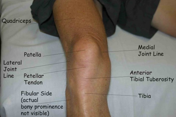
Anatomy Of The Left Knee Diagram
 www.mikrora.com
ucsd bursitis medial palpation tibial tibia meded patella muscle clergyman femur infrapatellar tuberosity
Anteroposterior Radiology (X-ray) Of The Knee : Anatomy Of The Femur
 www.pinterest.com
knee anatomy ray lower bones arteries xray extremity left anteroposterior radiology limb femur condyle medial normal ap atlas imaios lateral
198 Best Images About Chiropractic On Pinterest | Family Chiropractic
 www.pinterest.com
chiropractic knee
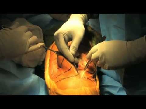
Total Knee Replacement Surgery 2011 - HD - YouTube
 www.youtube.com
knee replacement surgery total tkr happens explained expect pain there
Knee Posterior View
 www.thinglink.com
knee posterior
Radiographic Anatomy Of The Skeleton: Ankle -- Mortise View, Labelled
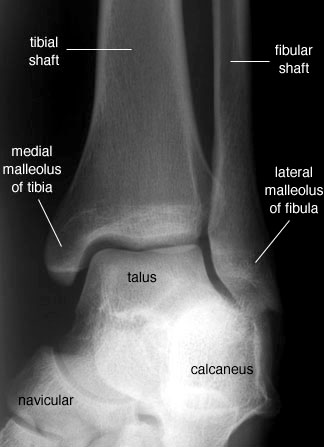 uwmsk.org
mortise ankle radiology anatomy labelled radiographic foot labeled lateral oblique medical talus ortopedia anatomical unlabelled version
Anatomy Of The Foot Medical Illustration Medivisuals
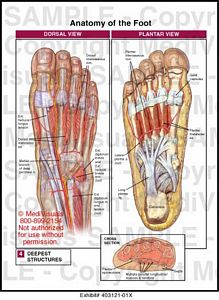 medivisuals1.com
foot anatomy 01x plantar dorsal right medical medivisuals1 illustration
CRACKCast E057 – Knee And Lower Leg - CanadiEM
 canadiem.org
crackcast e057 canadiem tibia
(hamstrings) biceps femoris: short head. Anteroposterior radiology (x-ray) of the knee : anatomy of the femur. Knee anatomy ray lower bones arteries xray extremity left anteroposterior radiology limb femur condyle medial normal ap atlas imaios lateral