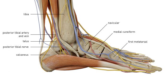
anterior ankle anatomy
Radiographic Anatomy of the Skeleton: Cervical Spine -- Right Anterior. 15 Pics about Radiographic Anatomy of the Skeleton: Cervical Spine -- Right Anterior : Ankle Anatomy - Sprain, Clinical Anatomy, Fracture, Radiology, X-Ray, google insight trending: Ankle injury and also Posterior ankle impingement | Image | Radiopaedia.org.
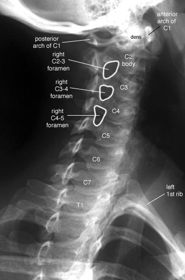
Radiographic Anatomy Of The Skeleton: Cervical Spine -- Right Anterior
 uwmsk.org
spine cervical anatomy oblique radiology ray anterior right labelled radiographs normal radiographic lumbar medical cspine student skeleton uwmsk imaging ap
Posterior Ankle Impingement | Image | Radiopaedia.org
 radiopaedia.org
ankle posterior impingement radiopaedia radiology process stieda lateral talar talus case posterolateral visit
Anterior View Of The Superficial Muscles Of The Leg | ClipArt ETC
 etc.usf.edu
leg muscles anterior superficial clipart etc lg usf edu
Ankle Ultrasound
 fpnotebook.com
ultrasound ankle anterior dorsal technique
Surface Anatomy Anterior View Of The African Amerian Foot | Joel Gordon
 joelgordon.photoshelter.com
surface anatomy foot anterior ics human
Rectus Capitis Posterior Minor: Origin, Insertion, Action | Kenhub
capitis rectus posterior minor muscle musculus kenhub insertion origin anatomy
Google Insight Trending: Ankle Injury
 google-insight-trending.blogspot.com
ankle anatomy injury google
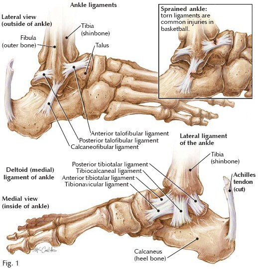
Sprained Ankle Anatomy - Human Anatomy Diagram | Ankle Anatomy
 www.pinterest.com
ankle ligaments anatomy human sprained diagram ligament tendons lateral muscle sprains foot bones sprain syndrome impingement management anterior treatment pain
Open And Arthroscopic Surgical Anatomy Of The Ankle
 www.hindawi.com
hardware arthroscopic fracture
Ankle Anatomy Health - Ankle Anatomyankle Anatomy
 greenhostit.com
ligaments tibia talus medial bones gland fibula adrenal greenhostit
The Anatomy Of The Posterior Ankle. Source: Norman, W. The Anatomy
 www.researchgate.net
posterior
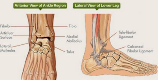
Ankle Anatomy - Sprain, Clinical Anatomy, Fracture, Radiology, X-Ray
 ehealthhall.com
ankle anatomy lateral pain anterior knee clinical running during joint ray radiology fracture squatting ankles while sprain courtesy
0514 Ankle Posterior Medical Images For Powerpoint | Presentation
 www.slideteam.net
posterior ankle powerpoint medical skip end
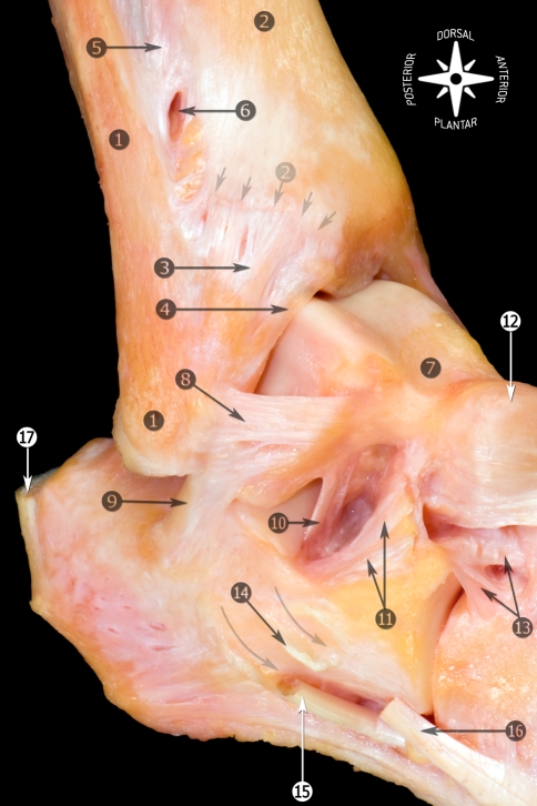
Ankle Ligaments - Foot & Ankle - Orthobullets
 www.orthobullets.com
ankle ligaments anatomy orthobullets foot lateral joint dissection ligament talofibular atfl collateral talocrural anterior pictorial essay calcaneofibular sprain dorsiflexion main
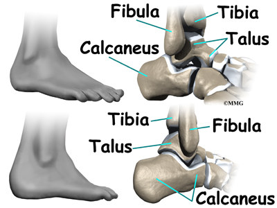
Ankle Anatomy | EOrthopod.com
 eorthopod.com
ankle anatomy bones foot lower joints leg plantar dorsal surface
Hardware arthroscopic fracture. Radiographic anatomy of the skeleton: cervical spine -- right anterior. Ultrasound ankle anterior dorsal technique