canine hock anatomy
Tarsal Anatomy of the Horse. 18 Images about Tarsal Anatomy of the Horse : Hock Anatomy Dog - Anatomy Drawing Diagram, The Equine Hock: What Horse Owners Should Know - Thal Equine LLC and also Hock Anatomy Dog.
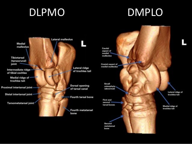
Tarsal Anatomy Of The Horse
 www.slideshare.net
tarsal metatarsal medial dorsal plantar
Steve Brooks K9U Steve's Articles - Steve Brooks K9U
 www.stevebrooksk9u.com
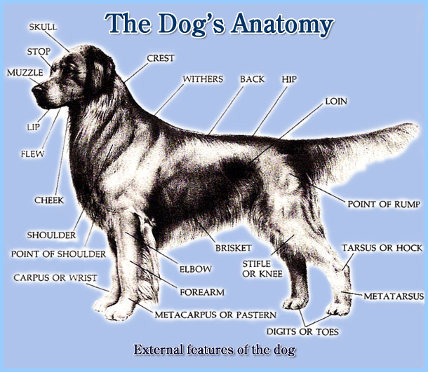
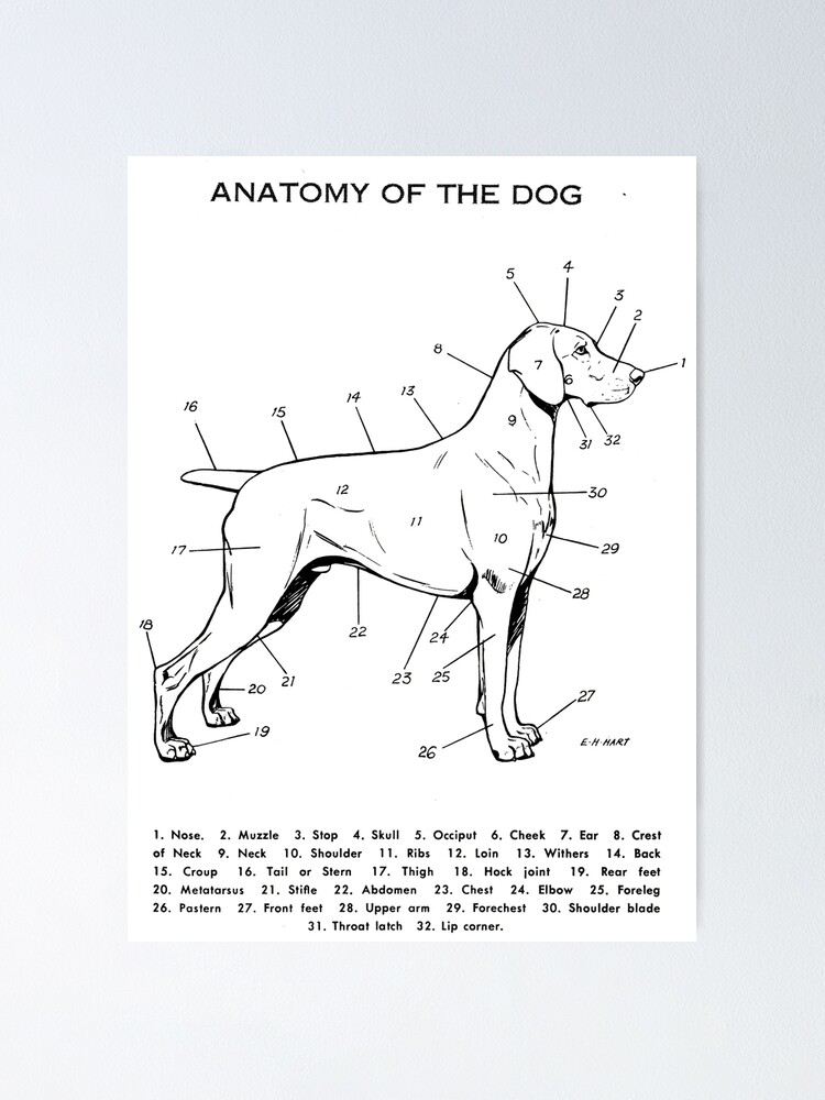
anatomy dog external articles
Canine Pelvic Limb Muscles Flashcards | Quizlet
 quizlet.com
canine muscles pelvic limb quizlet semitendinosus extend tibia flex stifle
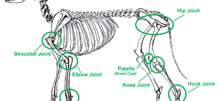
Hock Anatomy Dog - Anatomy Drawing Diagram
 sen842cova.blogspot.com
hock anatomy dog dogs diagram hindquarter drawing legs ankle joint
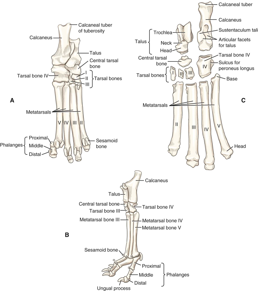
Canine Anatomy | Veterian Key
 veteriankey.com
anatomy canine hindlimb dog forelimb skeleton left hindpaw lateral plantar veterian key dorsal figure veteriankey
The Equine Hock: What Horse Owners Should Know - Thal Equine LLC
 www.pinterest.com
equine horses hock lameness
Pin By Overton Loyd On Dog Portraits | Dog Leg, Anatomy, Dog Anatomy
 www.pinterest.jp
carpus bones pasterns pastern skeletal fracture dislocation
GPI 9050 Canine Knee Model
 www.universalmedicalinc.com
knee canine anatomy joint dog vertebrae models veterinary move mouse enlarge anatomical gpi
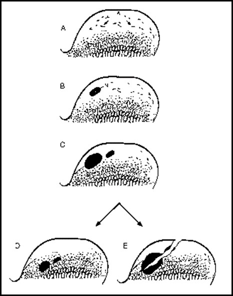
Hock Anatomy Dog
 greatbookfast.blogspot.com
hock ocd dissecans osteochondritis fitzpatrick referrals
Dog Anatomy Hock
 dogbreedsbest.blogspot.com
hock
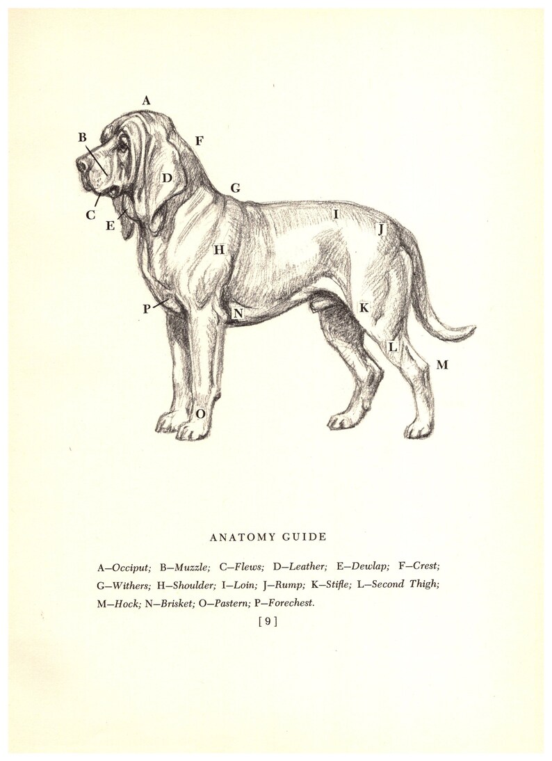
Hock Anatomy Dog
 greatbookfast.blogspot.com
anatomy hock megargee bloodhound edwin
Pin On Joint Health In Older Dogs
 www.pinterest.com
medial
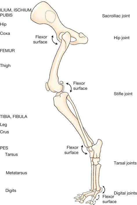
Canine Anatomy | Veterian Key
 veteriankey.com
anatomy canine hindlimb skeleton joints left dog flexor veterian key surfaces noting evans miller
Canine Anatomy Archives - Learn2GroomDogs
 www.learn2groomdogs.com
movement structure canine books
Canine Hindlimb Anatomy - Anatomy Drawing Diagram
 sen842cova.blogspot.com
thoracic hindlimb musculature limb subcutaneous medial muscles vetstudent dissection hindquarter
Illustration Of Parts Of A Dog - Google Search | Dog Anatomy, Dog Club
 www.pinterest.com
dog external anatomy parts collie grooming dogs anatomy2 bones project bearded drawing club records built
Dog Anatomy Hock
 dogbreedsbest.blogspot.com
hock
Ankle - TopDogHealth.com
 www.topdoghealth.com
ankle orthopedic knees dog where lower fracture surgery leg bone animal body hurting learn any condition
Dog external anatomy parts collie grooming dogs anatomy2 bones project bearded drawing club records built. Carpus bones pasterns pastern skeletal fracture dislocation. Canine pelvic limb muscles flashcards