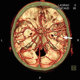
cerebral angiography anatomy
Bilateral caudate nucleus infarction associated with variant in circle. 18 Pics about Bilateral caudate nucleus infarction associated with variant in circle : Anatomy - Museum of the History of Science : Museum of the History of, What Is a Cerebral Angiography? and also Angiography (angiogram) adapted from angioCT showing all pelvic.
Bilateral Caudate Nucleus Infarction Associated With Variant In Circle
 casereports.bmj.com
infarction willis circle caudate cerebral artery anterior left a1 angiography figure mr bilateral bmj nucleus associated variant 2009 segment jnnp
Cerebral Angiography - Radiology Consultants
 radiology-consultants.com
cerebral angiography consultants radiology brain rays contrast flowing dye uses blood test
Cerebral Angiography - Dr Bassam - YouTube
 www.youtube.com
cerebral angiography
Cleveland Clinic Primary Angiitis Of The Central Nervous System
 teachmemedicine.org
angiitis central nervous angiography system primary cerebral
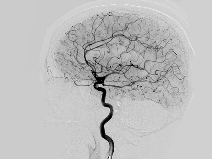
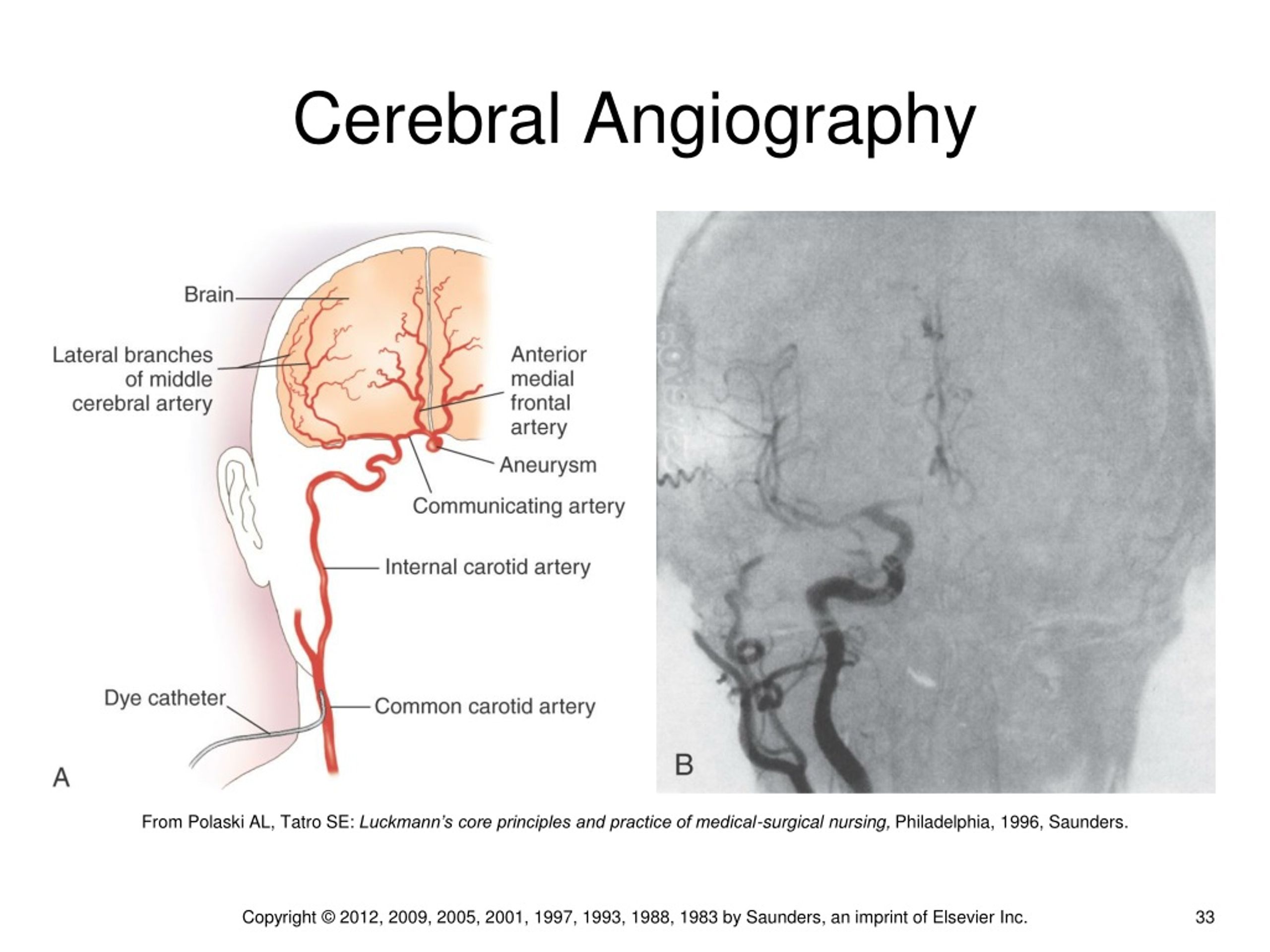
What Is A Cerebral Angiography?
 www.healthline.com
angiography dsa cuci catheterization otak healthline angiogram vessels terapi mengenal substraction diagnosis artery carotid
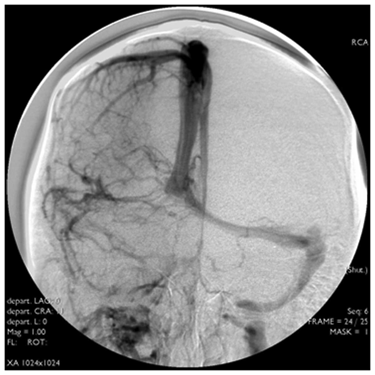
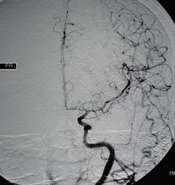
Early Imaging Characteristics Of 62 Cases Of Cerebral Venous Sinus
 www.spandidos-publications.com
sinus thrombosis venous cerebral dsa angiography subtraction digital lateral imaging characteristics cases early etm
Brain
 brainandspineclinic.com
stroke light brain symptoms getty
Cerebral Angiography | Radiology Key
 radiologykey.com
cerebral angiography fig
PPT - Chapter 15 PowerPoint Presentation, Free Download - ID:241128
 www.slideserve.com
chapter ppt powerpoint presentation
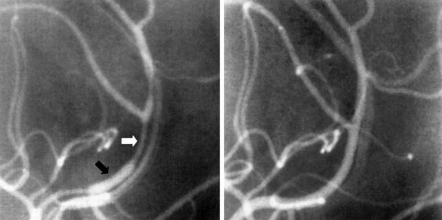
Artery Stenting: Basic Techniques | Thoracic Key
 thoracickey.com
artery angiography carotid stenting
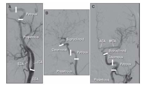
Cerebral Angiography | SpringerLink
 link.springer.com
cerebral angiography basilaris fig normal cerebri
PPT - Strokes In The Elderly PowerPoint Presentation, Free Download
 www.slideserve.com
elderly strokes ppt powerpoint presentation
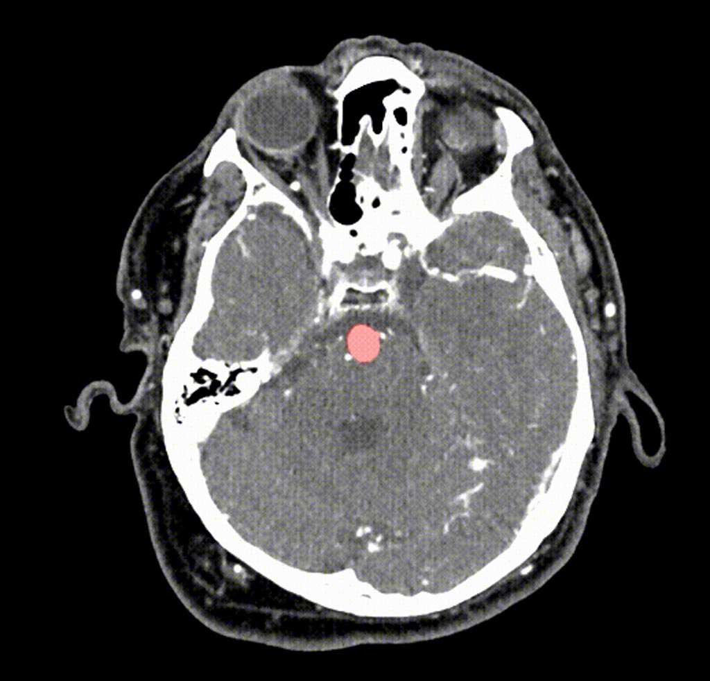
TradeMed - Industry News - New AI Tool Helps Detect Brain Aneurysms On
 www.trademed.com
brain ct scan angiography aneurysms ai aneurysm detect tool exams helps trademed stanford developed researchers university usa
Medical Sharing: Cerebral Angiography Indications
 medical75.blogspot.com
cerebral angiography indications angiogram sharing medical
Angiography (angiogram) Adapted From AngioCT Showing All Pelvic
 www.pinterest.com
anatomy arteries artery angiography pelvic angiogram labeled lower internal external pudendal extremity femoral aorta abdominal gluteal iliac leg limb label
Let's Recognize And Pay Homage To Portuguese Genius
 www.auntminnieeurope.com
cerebral angiography 1932 portuguese fig homage recognize genius pay let
Anatomy - Museum Of The History Of Science : Museum Of The History Of
 www.mhs.ox.ac.uk
anatomy brain mri willis circle angiogram radiology angio ac ox mhs screen cerebral human ανατομία branches humor rapid presentations aid
Radiology Images
 anatomy.elpaso.ttuhsc.edu
angiogram carotid internal superficial xray cerebral
Artery angiography carotid stenting. Angiography dsa cuci catheterization otak healthline angiogram vessels terapi mengenal substraction diagnosis artery carotid. Angiography (angiogram) adapted from angioct showing all pelvic