ct scan brain anatomy
Sagittal midline of the brain: normal anatomy | Radiology Case. 17 Pictures about Sagittal midline of the brain: normal anatomy | Radiology Case : Normal anatomy of the brain on CT and MRI with a few normal variants, Anatomy Of Brain Ct Scan - Anatomy Drawing Diagram and also Basics of brain CT scan part III - YouTube.
Sagittal Midline Of The Brain: Normal Anatomy | Radiology Case
 www.pinterest.com
cisterns radiology radiopaedia mri sagittal normal venous midline subarachnoid cistern cerebral galen intracranial malformations quadrigeminal cisterna prepontine terminalis csf interpeduncular
CT Brain With Severe Motion Artifact | Image | Radiopaedia.org
 www.radiopaedia.org
ct artifact motion brain severe artifacts movement radiopaedia case patient version based
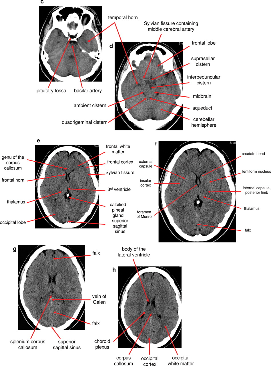
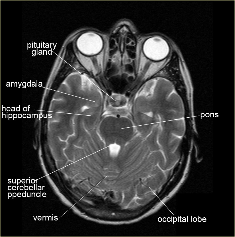
Normal Anatomy Of The Brain On CT And MRI With A Few Normal Variants
 pn.bmj.com
ct anatomy brain scan normal head mri powerpoint cranial figure tab open info
Brain Jack Image: Brain Ct Scan
 brainjackimage.blogspot.com
ct scan normal brain scans head anatomy medical human acute crash radiology shift ctscan jack someone university international addicts case
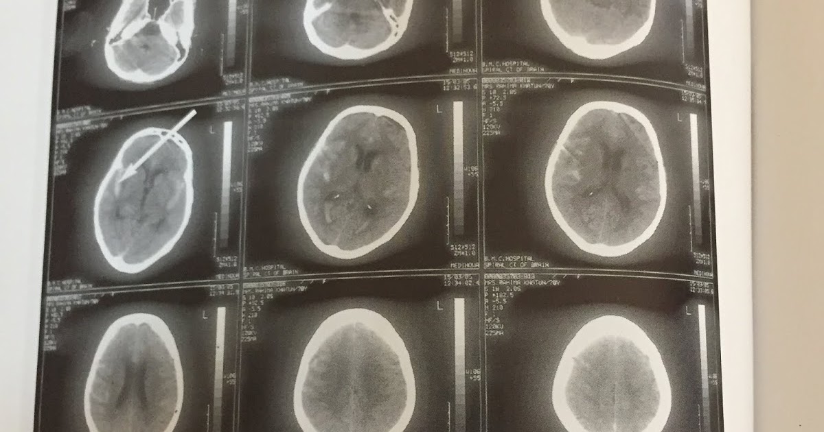
CT SCAN OF BRAIN-6
 medicalschoolimportant.blogspot.com
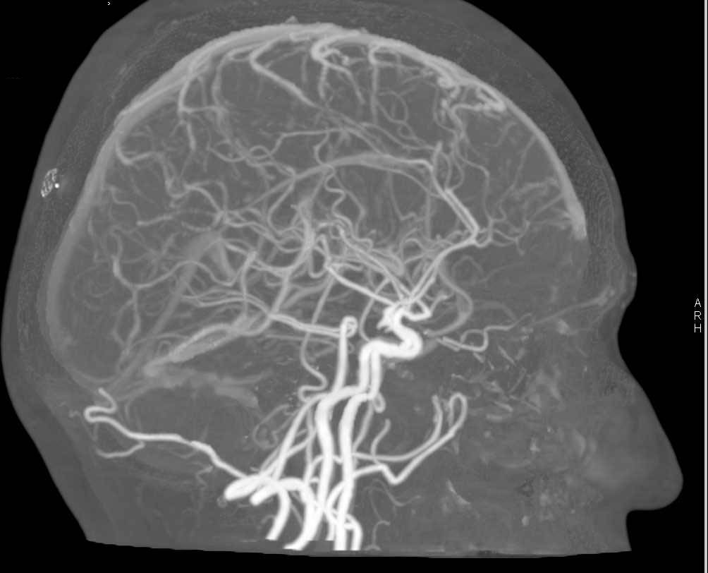
CT Scan Of The Brain Vessels - YouTube
 www.youtube.com
scan brain
Ct Scan Brain Anatomy : Anatomy Of Head Ct Scan Normal The Brain On Ct
 saripepaya11.blogspot.com
ct axial radiology labelled cerebral pituitary gland lobe cortex radiologie transverse radiologyassistant neurology assistant cerebro radiographic medizin basal neuroscience orbits
Axial And Helical Ct Scans - Ct Scan Machine
 ctscanmachines.blogspot.com
helical scans
Anatomy Of Brain Ct Scan - Anatomy Drawing Diagram
 sen842cova.blogspot.com
basal ganglia radiology axial neuroanatomy diagram transverse nuclei subthalamic radiologyassistant 神経 anatomical 科学 人体
Basics Of Brain CT Scan Part III - YouTube
 www.youtube.com
CTA Of Circle Of Willis - Neuro Case Studies - CTisus CT Scanning
 www.ctisus.com
ct cta willis circle brain ctisus scan neuro removal bone studies case diagnosis
Brain Jack Image: Brain Ct Scan
 brainjackimage.blogspot.com
brain ct scan perfusion scans sinai imaging jack cedars usi scan3 use
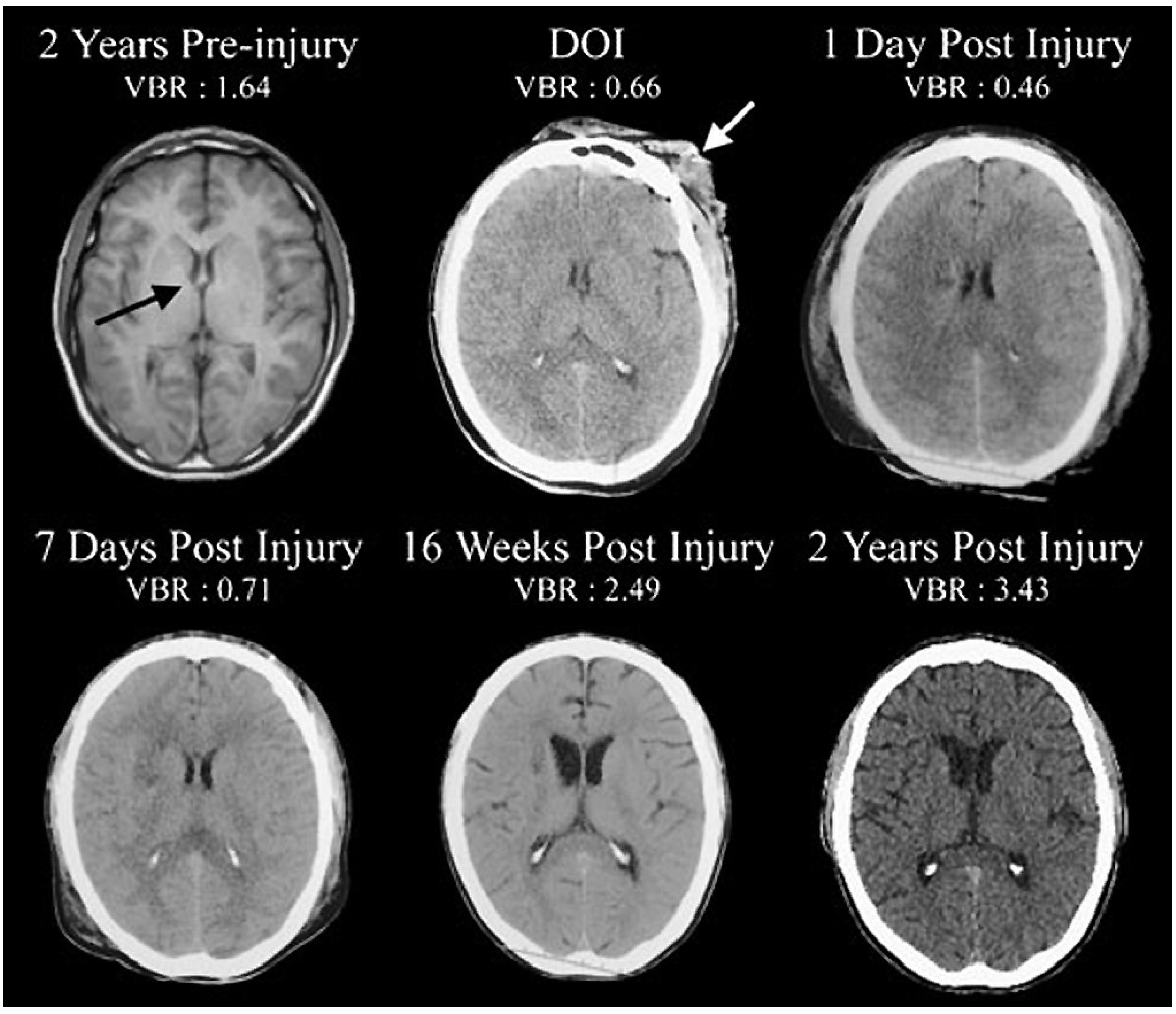
Brain Sciences | Free Full-Text | Damage To Myelin And Oligodendrocytes
 www.mdpi.com
brain injury traumatic damage mri tbi myelin imaging severe years brainsci prior chronic outcomes oligodendrocytes role following mdpi
Malignant Middle Cerebral Artery (MCA) Infarction: Pathophysiology
 pmj.bmj.com
ct herniation uncal mca artery cerebral middle left infarction medical malignant figure 1014 bmj pmj
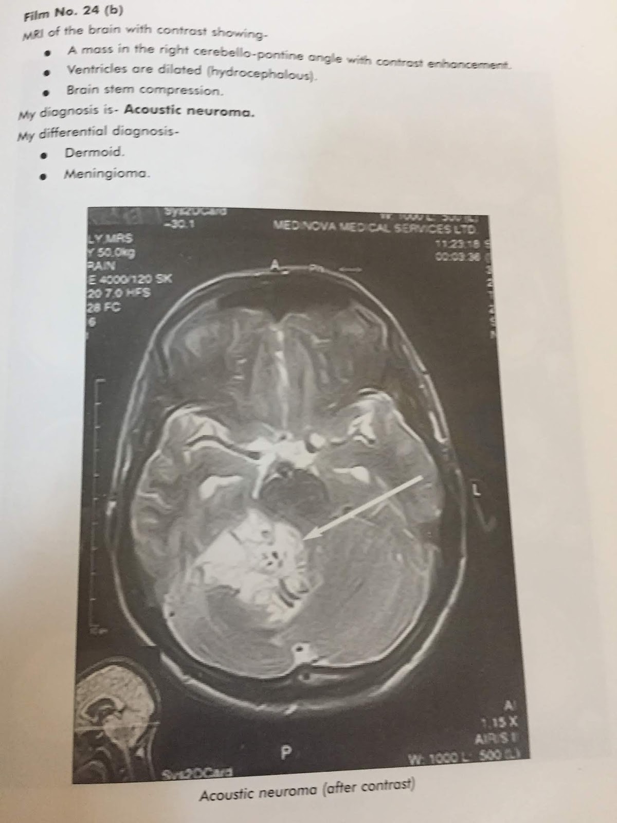
CT SCAN OF BRAIN-14
 medicalschoolimportant.blogspot.com
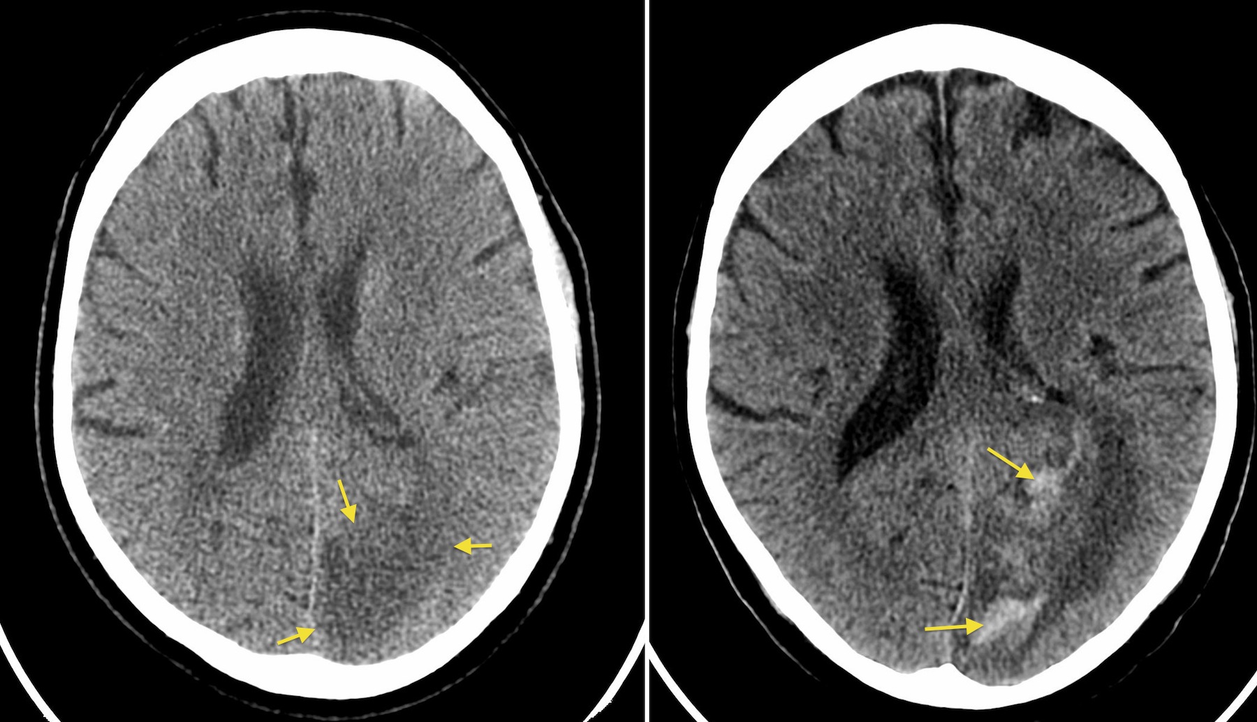
Haemorrhagic Transformation Of CVA - Radiology At St. Vincent's
 www.svuhradiology.ie
cva transformation haemorrhagic radiology svuhradiology ie
Posterior Fossa Vascular Territories | Image | Radiopaedia.org
 radiopaedia.org
posterior inferior fossa cerebellar artery vascular territories radiopaedia axial cerebellum pica radiology brain right infarct case version cranial
Ct scan of brain-6. Brain jack image: brain ct scan. Brain sciences