dental radiographic anatomy
PPT - EO 005.06 Normal Intraoral Radiographic Anatomy PowerPoint. 17 Images about PPT - EO 005.06 Normal Intraoral Radiographic Anatomy PowerPoint : Radiographic Anatomy on Pinterest | Anatomy, Dental Anatomy and Bones, Dental Radiographic Anatomy - YouTube and also 33: Extraoral Radiographic Landmarks | Pocket Dentistry.
PPT - EO 005.06 Normal Intraoral Radiographic Anatomy PowerPoint
 www.slideserve.com
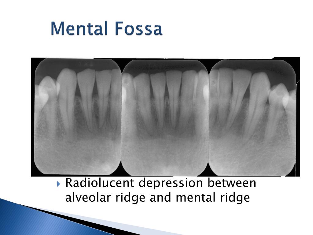
mental fossa radiolucent ridge intraoral radiographic eo anatomy normal ppt powerpoint presentation alveolar depression between
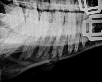
Anatomy – Hamulus – Pantomograph – Drgstoothpix – Dr. G's Toothpix
 drgstoothpix.com
hamulus anatomy radiopaque tuberosity maxillary drgstoothpix side right monday entity distal showing
Enamel Pearl – Dr. G's Toothpix
 drgstoothpix.com
enamel pearl tooth molar radiopaque third radiographic roots note round area interpretation
Radiographic Anatomy On Pinterest | Anatomy, Dental Anatomy And Bones
 www.pinterest.com
radiography radiographic maxillofacial mandible panoramic hygiene
Advances In Radiographic Techniques Used In Dentistry | IntechOpen
 www.intechopen.com
radiographic dentistry advances techniques intechopen figure
PPT - Extraoral Radiographic Anatomy PowerPoint Presentation, Free
 www.slideserve.com
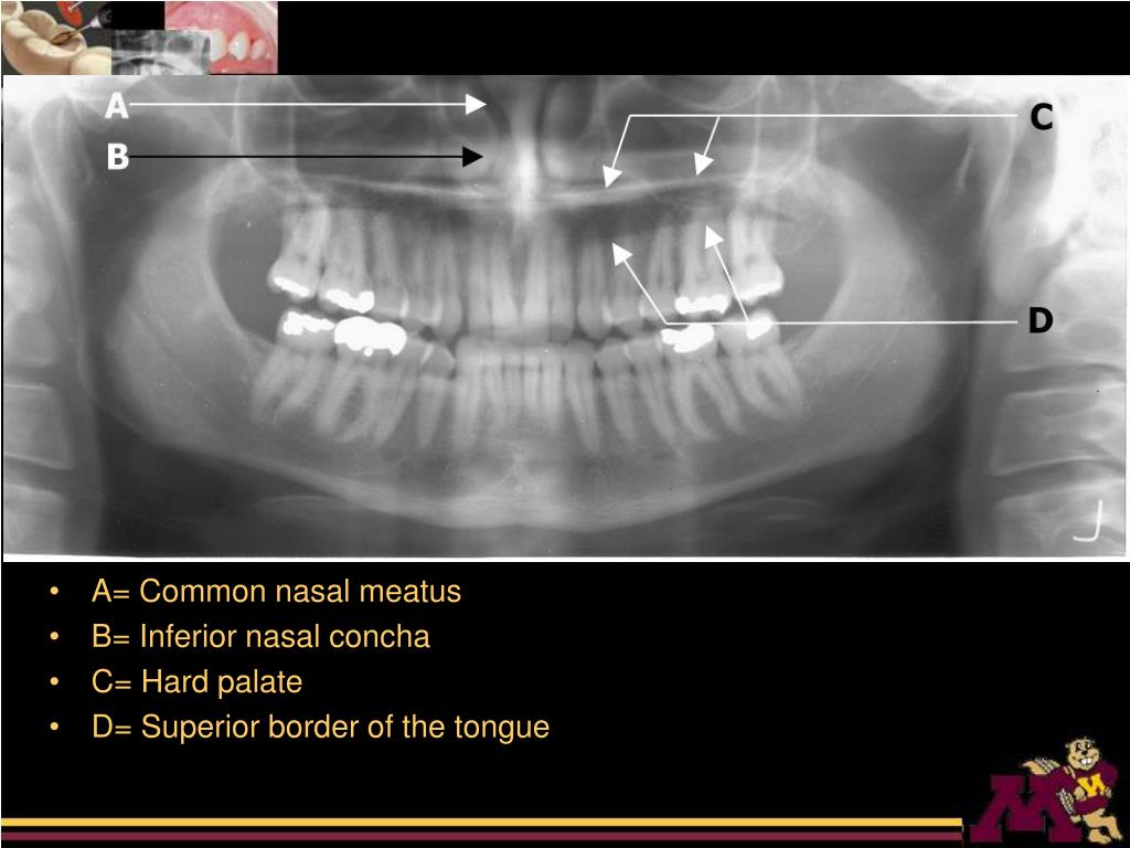
nasal inferior concha extraoral anatomy superior process palate hard radiographic rim meatus common tongue border
Dental, Anatomy And Finals On Pinterest
 www.pinterest.com
dental landmarks radiographic anatomy mandibular pano radiology oral assistant study hygiene ray dentistry finals structures radiograph panoramic anatomical help hygienist
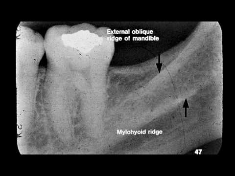
33: Extraoral Radiographic Landmarks | Pocket Dentistry
 pocketdentistry.com
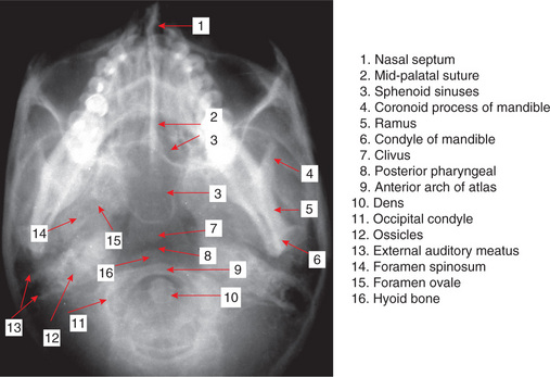
landmarks radiographic extraoral figure pocketdentistry
Radiology
 www.equerry.ca
radiology
Dental Radiographic Interpretation: 1. Normal Anatomy - YouTube
 www.youtube.com
Dental Radiographic Anatomy - YouTube
 www.youtube.com
dental anatomy radiographic oral radiograph landmarks radiology tooth teeth panoramic intraoral structures bridge
1 Dental Radiology
 www.slideshare.net
radiology
The Art Of Dental Radiography - Dimensions Of Dental Hygiene
 dimensionsofdentalhygiene.com
radiography
Radiographic Interpretation
 www.slideshare.net
radiographic opg anatomy describing lesion
Dental Assistant Radiology Study Guide
 thetancollective.com
dermoid radiology ruptured radiopaedia arachnoid sub bleed subarachnoid brain mri cyst ct radio case study dental guide hemorrhage imaging lobe
DENTAL RADIOGRAPHY - Principles And Techniques. | Vebuka.com
 vebuka.com
Panoramic Radiography — Diagnosis Of Relevant Structures That Might
 www.intechopen.com
panoramic radiography oral general structures diagnosis health compromise relevant might intechopen figure patient
Anatomy – hamulus – pantomograph – drgstoothpix – dr. g's toothpix. 33: extraoral radiographic landmarks. Enamel pearl – dr. g's toothpix