equine tarsal anatomy
PPT - Normal Radiographic Anatomy of the Equine Hind Limb PowerPoint. 16 Images about PPT - Normal Radiographic Anatomy of the Equine Hind Limb PowerPoint : Anatomy and Radiology on Pinterest, Equine hock oblique posterior view and also PPT - Normal Radiographic Anatomy of the Equine Hind Limb PowerPoint.
PPT - Normal Radiographic Anatomy Of The Equine Hind Limb PowerPoint
 www.slideserve.com
equine cont tarsus
Equine Carpus Anatomy
 www.slideshare.net
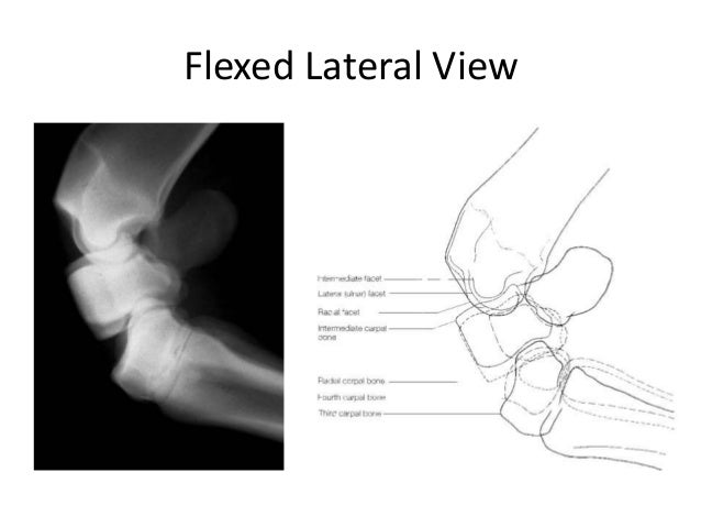
carpus flexed anatomy carpal distal
Equine Hock Oblique Posterior View
 inkymousestudios.com
hock equine oblique posterior joint project
Equine Carpus Anatomy
 www.slideshare.net
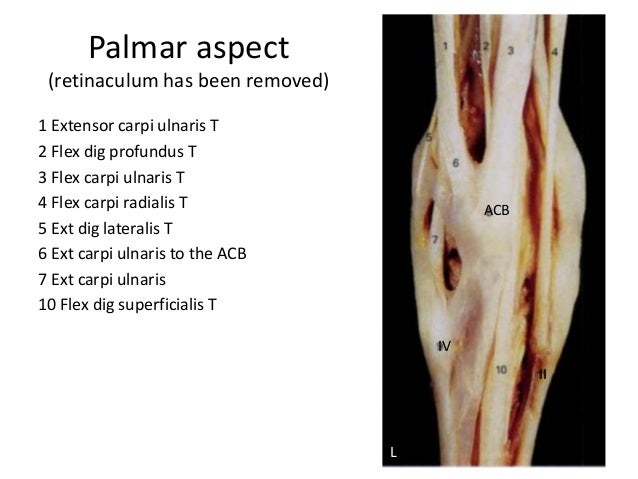
carpi equine retinaculum extensor palmar carpus carpal ulnaris
Anatomy Of The Horse
 www.slideshare.net
www.slideshare.net
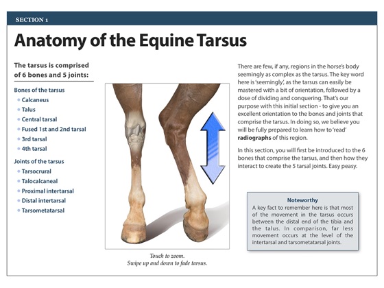
tarsal
Anatomy Arthrology Flashcards | Quizlet
 quizlet.com
anatomy horse tarsus arthrology joint quizlet
Equine Tarsal Bones Quiz
 www.purposegames.com
PPT - Normal Radiographic Anatomy Of The Equine Hind Limb PowerPoint
 www.slideserve.com
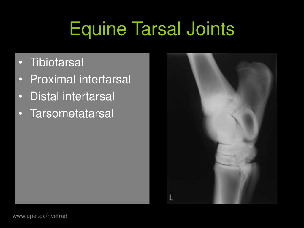
equine tarsal joints intertarsal distal radiographic hind limb anatomy normal proximal tarsometatarsal upei tibia ppt powerpoint presentation
Disorders Of The Tarsus In Horses: Bone, Joint, And Muscle Disorders In
 www.pinterest.jp
anatomy horse hock muscle disorders horses tarsus health equine joint leg pet bone veterinary manual merck anatomia tendons medicine diagram
Anatomy And Radiology On Pinterest
 www.pinterest.com
anatomy horse equine radiology hock veterinary anatomie care pferd vet tarsus dog animal medicine joint tech joints
Tarsal Anatomy Of The Horse
 www.slideshare.net
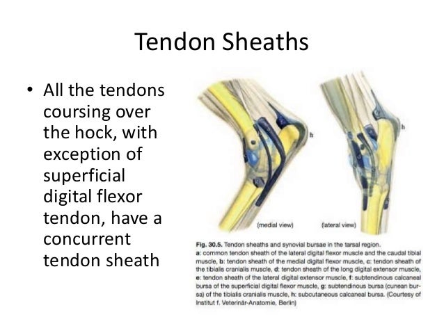
tarsal hock tendon tendons ligament sheaths medial collateral malleolus
Anatomy And Radiography Of The Equine Tarsus On Apple Books
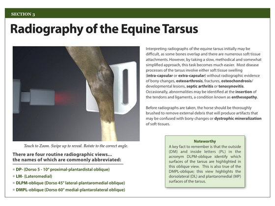 books.apple.com
tarsus radiography
Astragalo Stock Photos & Astragalo Stock Images - Alamy
 www.alamy.com
astragalo domesticated comparative horses
Gluteobiceps - Animal Anatomy - Joshua Nava Arts
 www.joshuanava.biz
anatomy animal calcaneus equine outside joshua nava arts
Anatomy And Radiography Of The Equine Tarsus On Apple Books
 books.apple.com
tarsus anatomy equine horse radiography bones
Anatomy II Lab - Midterm (equine) Flashcards - Cram.com
 www.cram.com
fossa extensor femur equine midterm anatomy lab ii flashcards cram
anatomy and radiography of the equine tarsus on apple books. anatomy and radiography of the equine tarsus on apple books. Disorders of the tarsus in horses: bone, joint, and muscle disorders in