foot x ray anatomy
Foreign body in foot | Image | Radiopaedia.org. 17 Images about Foreign body in foot | Image | Radiopaedia.org : Radiographic Anatomy - Foot DP | Life As I Know It | Pinterest, Foot Bones X Ray / Cureus Chondromyxoid Fibroma Of Distal Phalanx Of and also Radiographic Anatomy - Foot DP | Life As I Know It | Pinterest.
Foreign Body In Foot | Image | Radiopaedia.org
 radiopaedia.org
radiopaedia
Tarsal Navicular Fracture | Image | Radiopaedia.org
 radiopaedia.org
navicular fracture tarsal radiopaedia oblique
Pin On Radiographic Anatomy
 www.pinterest.com.au
radiographic radiograph radiology bearing muscles radiografia wikiradiography physiology medial schools radiologic anatomia talus positioning radiography radiological
The Charcot Foot In Diabetes | Diabetes Care
 care.diabetesjournals.org
charcot foot diabetes care figure
Foot Bones X Ray / Cureus Chondromyxoid Fibroma Of Distal Phalanx Of
 lucindaj-sand.blogspot.com
Foot X Ray Anatomy
 savecatchingfire.blogspot.com
podiatry
Ankle Fracture - Weber B | Image | Radiopaedia.org
 radiopaedia.org
fracture weber ankle radiopaedia version joint
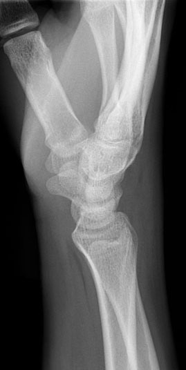
Radiographic Anatomy Of The Skeleton: Wrist -- Lateral View, Unlabelled
 uwmsk.org
wrist lateral anatomy lat ray xray xr labelled skeleton rays unlabelled version hover
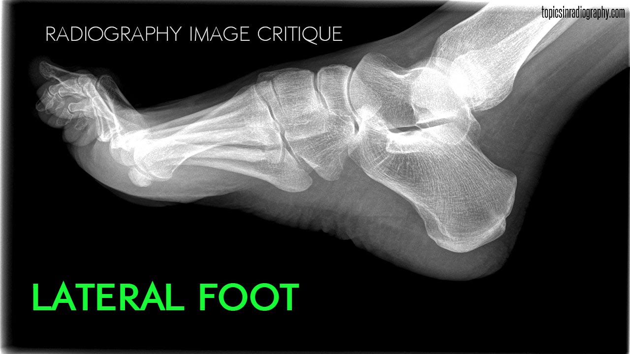
Topics In Radiography - YouTube
 www.youtube.com
Radiology Anatomy Images : Foot Lateral X-ray Anatomy
 radiology-anatomy.blogspot.com
anatomy
Radiographic Anatomy - Foot DP | Life As I Know It | Pinterest
 pinterest.com
foot anatomy radiographic wikiradiography ray bones labeled normal ap found views oblique right bone
Os Trigonum | Image | Radiopaedia.org
 radiopaedia.org
trigonum os radiopaedia lateral talus fracture posterior radio radiology foot mri case
Foot X-ray - Normal Findings | Bone And Spine
 boneandspine.com
foot xray normal ray bone bones findings radiology spine anatomy marked showing virginia boneandspine
Anatomy Xray Of The Foot. Learn More About Structural Anatomy And How
 www.pinterest.com
xray anatomy yin training yoga structural foot learn teacher
Student Study Guides - Foot Anatomy
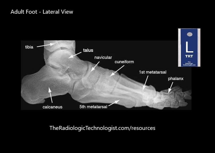 theradiologictechnologist.com
missing
Foot: Annotated X-ray | Image | Radiopaedia.org
 radiopaedia.org
foot ray radiograph annotated radiopaedia normal radiology metatarsals oblique approach phalanges radiographs figure version
Radiographic Anatomy - Foot Oblique. | Radiology Student, Radiology
 www.pinterest.com
foot anatomy bones oblique radiology
Navicular fracture tarsal radiopaedia oblique. Radiographic radiograph radiology bearing muscles radiografia wikiradiography physiology medial schools radiologic anatomia talus positioning radiography radiological. Radiology anatomy images : foot lateral x-ray anatomy