hip ultrasound anatomy
How to scan protocol - step by step FREE download. 17 Pics about How to scan protocol - step by step FREE download : How to scan protocol - step by step FREE download, The Ultrasound Board Review Book is Out! Exclusive POCUS 101 Discount and also Wrist Case 2 - Sports Medicine Imaging.
How To Scan Protocol - Step By Step FREE Download
 theultrasoundsite.co.uk
hip ultrasound joint protocol recess region
MRI Anatomy Of Hip Joint | Free MRI Axial Hip Anatomy In 2021 | Hip
 www.pinterest.com
hip anatomy axial mri joint cross sectional pelvis muscle section tensor sartorius latae use nerve sciatic fasciae move
Shoulder Anatomy On Ultrasound | Image | Radiopaedia.org
 radiopaedia.org
ultrasound anatomy shoulder radiopaedia rotator cuff radiology supraspinatus normal sonography case msk tendon muscle infraspinatus bursa muscles joint tendons teres
Hip Ultrasound Diagnostic Imaging | Melbourne Radiology
 www.melbourneradiology.com.au
undergo mri melbourneradiology
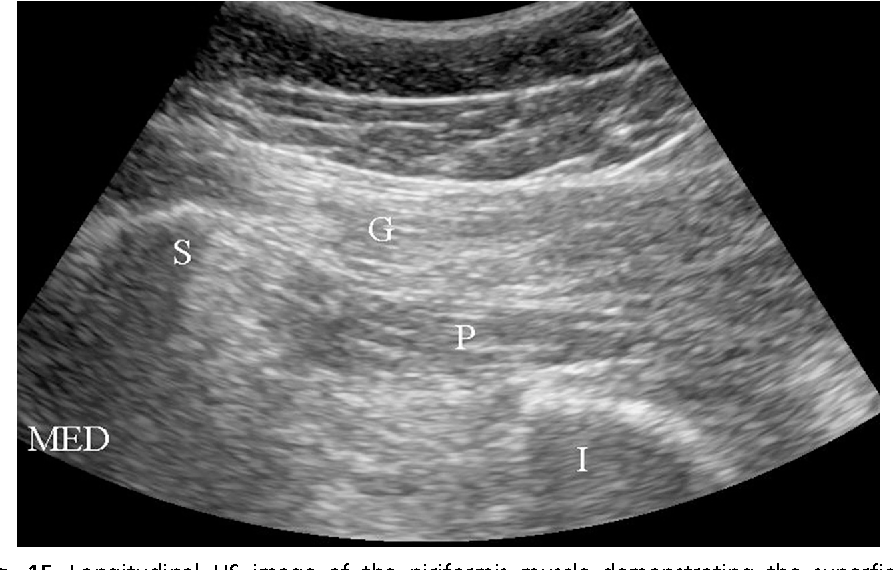
Ultrasound Of The Hip | SpringerLink
 link.springer.com
fig ultrasound hip
Image | Radiopaedia.org
 radiopaedia.org
effusion radiopaedia modality
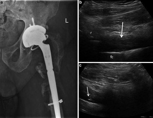
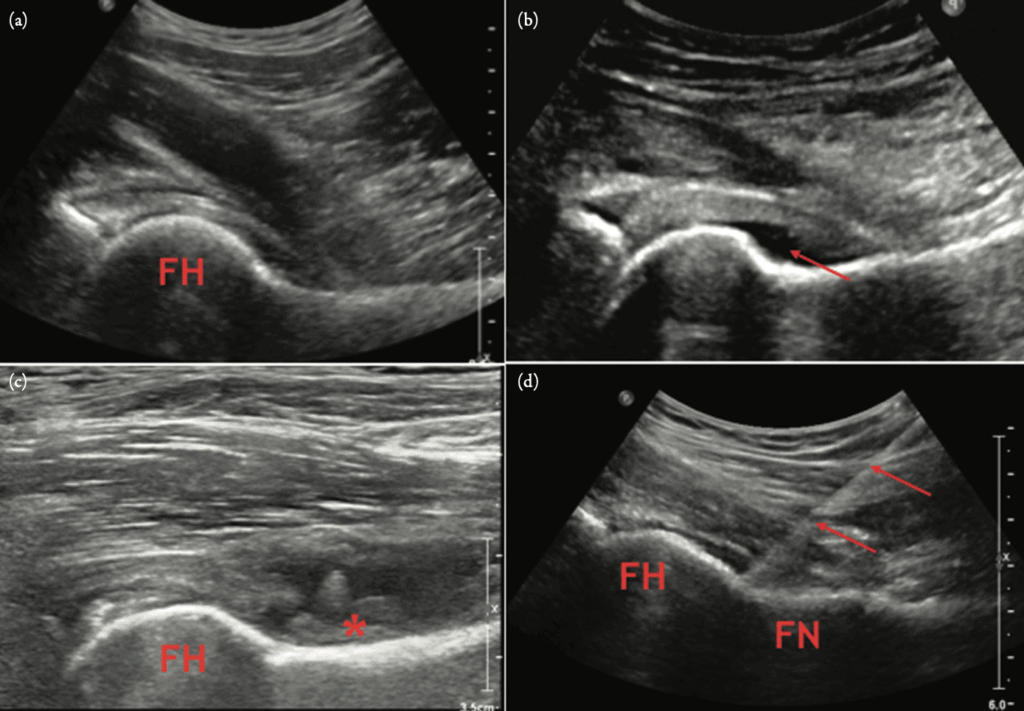
Figure 5 From Ultrasound-Guided Hip Procedures. | Semantic Scholar
 www.semanticscholar.org
ultrasound hip
Wrist Case 2 - Sports Medicine Imaging
 sportsmedicineimaging.com
arthrofibrosis
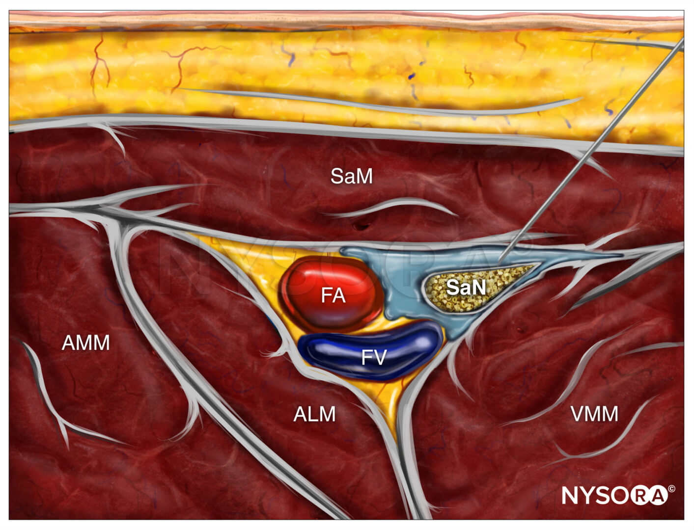
Ultrasound-Guided Saphenous (Adductor Canal) Nerve Block - NYSORA
 www.nysora.com
adductor saphenous nysora block canal nerve ultrasound guided anesthesia extremity lower
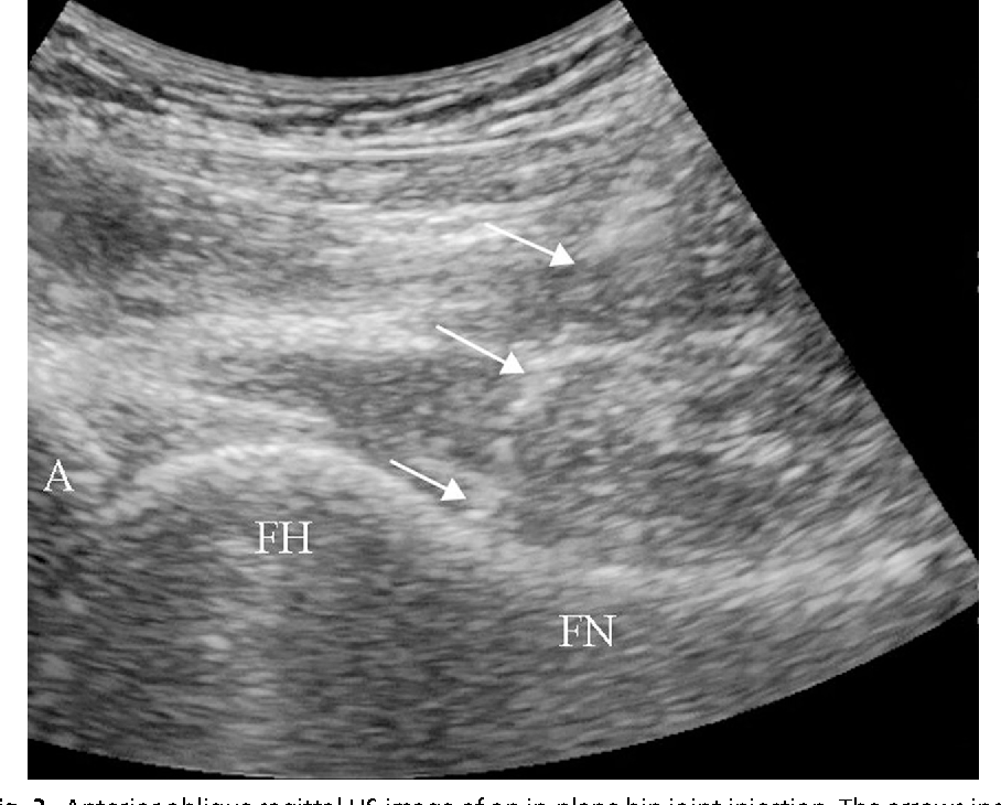
Figure 3 From Ultrasound-Guided Hip Procedures. | Semantic Scholar
 www.semanticscholar.org
ultrasound hip guided
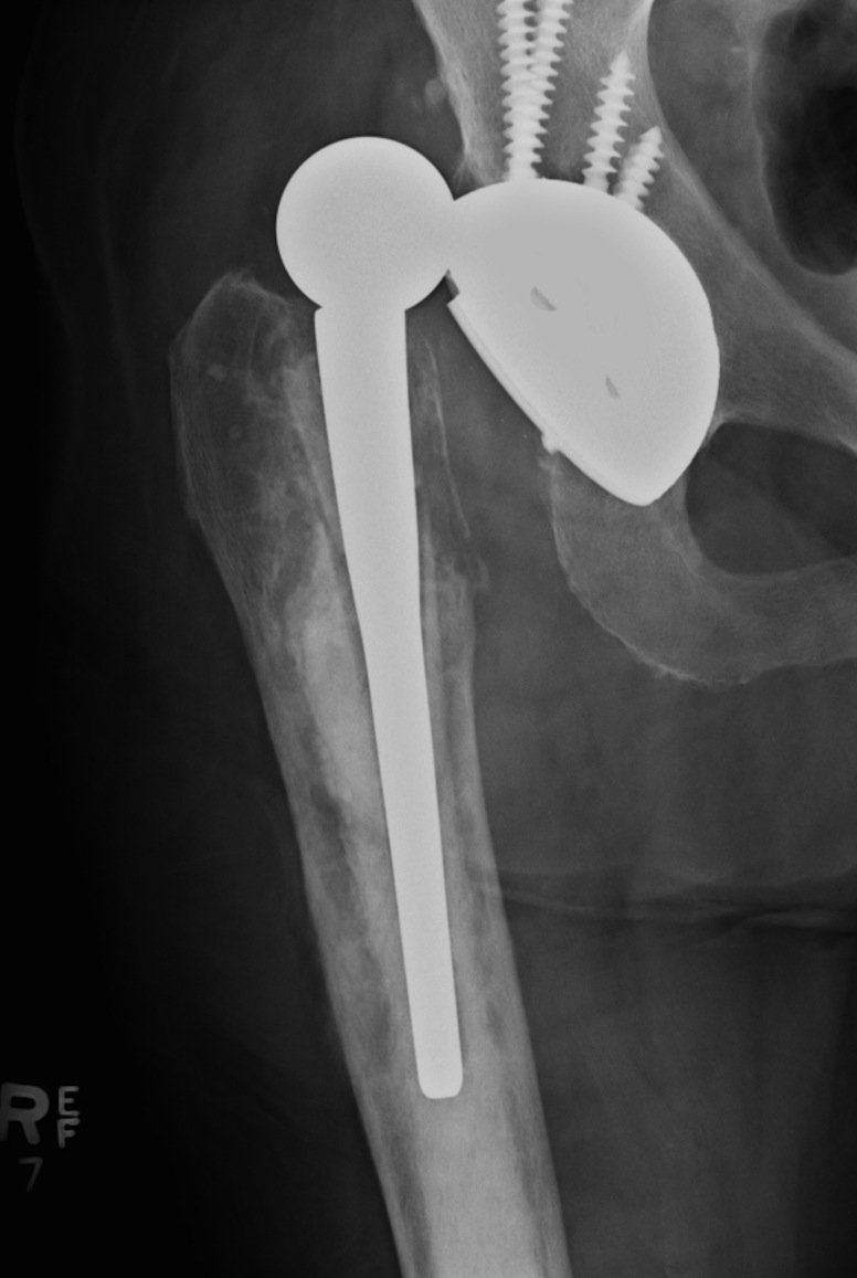
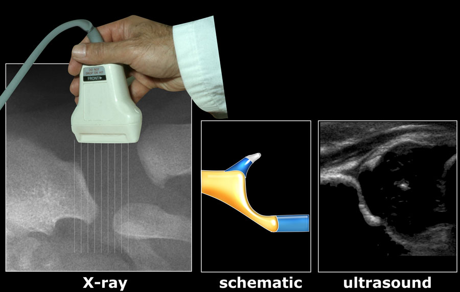
Dislocated Hip Prosthesis - Radiology At St. Vincent's University Hospital
 www.svuhradiology.ie
hip prosthesis
The Hip Joint - Complete Physiotherapy
 www.completephysiotherapy.co.uk
hip joint anatomy human bones body replacement ball resurfacing bone pelvis femur skeletal where system virtual diagram ligament spine conditions
Figure 14 From Ultrasound-Guided Hip Procedures. | Semantic Scholar
 www.semanticscholar.org
procedures
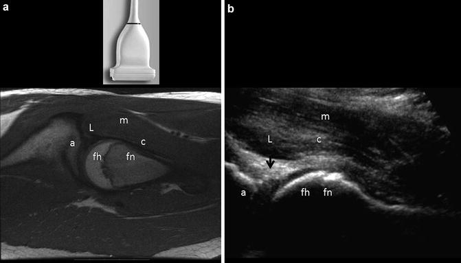
Ultrasound Of The Hip | SpringerLink
 link.springer.com
ultrasound hip fig iliopsoas
Hand Case 2 - Sports Medicine Imaging
 sportsmedicineimaging.com
hand stener case sports imaging
Anatomy For Ultrasound - Anatomy Diagram Book
 grekoulas.blogspot.com
ultrasound dysplasia developmental radiology
The Ultrasound Board Review Book Is Out! Exclusive POCUS 101 Discount
 www.pocus101.com
ultrasound pocus101 pocus
Ultrasound hip guided. Hand stener case sports imaging. The hip joint