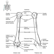
humerus x ray anatomy
Normal shoulder | Image | Radiopaedia.org. 16 Images about Normal shoulder | Image | Radiopaedia.org : Radiographic positioning: Humerus AP and lateral, 155 best Radiographic Anatomy images on Pinterest and also Humerus | Radiology Reference Article | Radiopaedia.org.
Normal Shoulder | Image | Radiopaedia.org
 radiopaedia.org
shoulder normal rays ray rotation internal external radiopaedia views version case roll zoom
Radiographic Positioning: Humerus AP And Lateral
 radiox-ray.blogspot.jp
humerus lateral anatomy radiology radiographic ray labeled student ap radiography positioning shoulder medical technology trauma xray tech hand science schools
155 Best Radiographic Anatomy Images On Pinterest
 www.pinterest.com
radiographic anatomy radiology
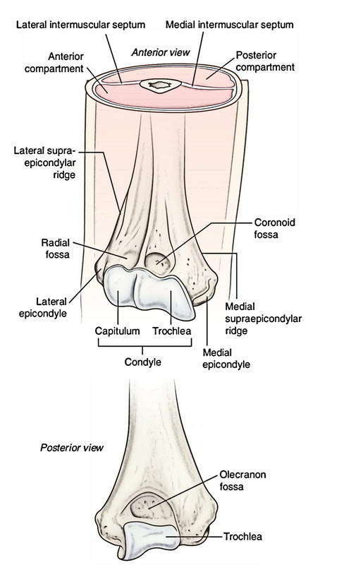
Osteology Of The Upper Limb - Last's Anatomy: Regional And Applied
 doctorlib.info
anatomy applied regional last figure lasts
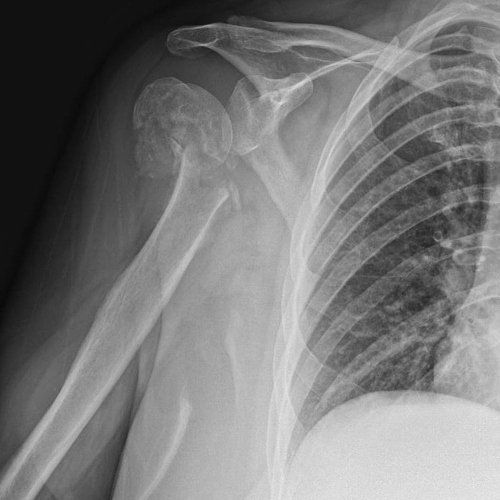
Surgical Neck Of Humerus Fracture | Image | Radiopaedia.org
 radiopaedia.org
fracture humerus neck surgical humeral proximal fractures radiology transverse radiopaedia case frontal simple non
Reverse Total Shoulder Replacement | Dr Sunil Reddy
 drsunilreddy.com.au
shoulder reverse replacement total upper limb fractures fracture humerus ray tuberosity proximal arthroplasty example reconstruction severely comminuted ct 3d
Proximal Humerus Anatomy | Humerus Anatomy, Anatomy, Physiology
 www.pinterest.com
anatomy humerus proximal physiology
Humerus - Anatomy QA
 www.anatomyqa.com
humerus anatomy
Proximal Humeral Fracture | Image | Radiopaedia.org
 radiopaedia.org
humeral proximal fracture fractures case radiopaedia radiology classification version fr loading
Humerus | Radiology Reference Article | Radiopaedia.org
 radiopaedia.org
humerus radiopaedia radiology figure figures cases
Supracondylar Fracture | Image | Radiopaedia.org
 radiopaedia.org
fracture elbow supracondylar pediatric radiopaedia fractures gartland classification humeral radiograph type humerus paediatric radiology capitellum cases radiographs line case orthopedics
Anteroposterior Radiology (X-ray) Of The Knee : Anatomy Of The Femur
 www.pinterest.com
knee anatomy ray lower bones arteries xray extremity left anteroposterior radiology limb femur condyle medial normal ap atlas imaios lateral
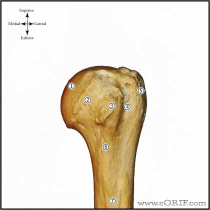
Proximal Humerus Anatomy
 eorif.com
humerus proximal anatomy bone shoulder head humeral neck tuberosity greater major groove lesser anterior pectoralis muscle anatomic eorif attachments bony
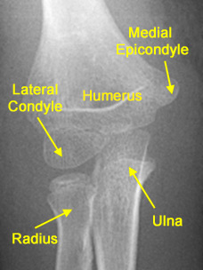
OrthoKids - Elbow Fractures
 orthokids.org
elbow bone fractures pediatric forearm
Humerus
 www.earthslab.com
humerus anatomy proximal end shaft distal fracture upper head
Supracondylar Fracture | Image | Radiopaedia.org
 radiopaedia.org
fracture elbow supracondylar fractures children pediatric radiopaedia complications obese likely complex frontal case version non
Osteology of the upper limb. Anatomy applied regional last figure lasts. Proximal humerus anatomy