knee anatomy x ray
my xray. 18 Pics about my xray : Normal Knee X-rays | Radiology student, Radiology schools, Medical anatomy, Dennis M. Lox, M.D. Explains How to Read a "Bone on Bone" Knee X-Ray and also The Radiology Assistant : Ankle - Special fracture cases.
My Xray
 www.kneeguru.co.uk
diagram xray ive borrowed marked pain orange where knee
Lipohaemarthrosis | Image | Radiopaedia.org
 radiopaedia.org
radiopaedia version case
X-Ray Picture Showing Knee Joints Stock Photo 254809927 : Shutterstock
 www.shutterstock.com
joints knee ray showing shutterstock
Interpreting X-Rays Of The Knee Joint - YouTube
 www.youtube.com
knee joint rays cyk
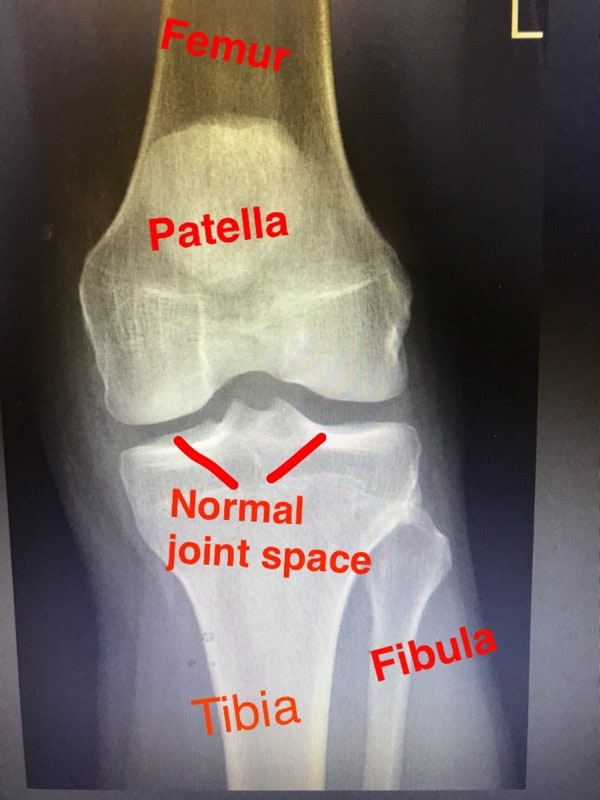
Dennis M. Lox, M.D. Explains How To Read A "Bone On Bone" Knee X-Ray
 www.drlox.com
knee ray normal bone read space joint
Fraturas | CTO
 cto.med.br
joelho patela fraturas fratura tibia joelhos cto
ACL Surgeon Adelaide | Knee Ligament Reconstruction Adelaide
 drsunilreddy.com.au
acl reconstruction knee normal arthroscopic ligament surgery
Vertical Fracture Of Patella | Image | Radiopaedia.org
 radiopaedia.org
fracture knee patella vertical cap radiopaedia version case
Anatomy Knee Xray - Human Anatomy
 tartrerepub.blogspot.com
xray radiopaedia
Bipartite Patella | Image | Radiopaedia.org
 radiopaedia.org
patella bipartite radiopaedia frontal
Normal Knee X-rays | Radiology Case | Radiopaedia.org
 radiopaedia.org
radiopaedia xray buyxraysonline crossovers
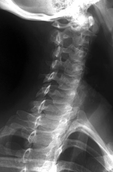
Radiographic Anatomy Of The Skeleton: Cervical Spine -- Right Anterior
 uwmsk.org
spine cervical anatomy oblique radiology ray anterior right radiographs labelled normal radiographic lumbar medical cspine student skeleton uwmsk imaging ap
Knee Anatomy
 mysurgery.nshealth.ca
knee joint anatomy bones
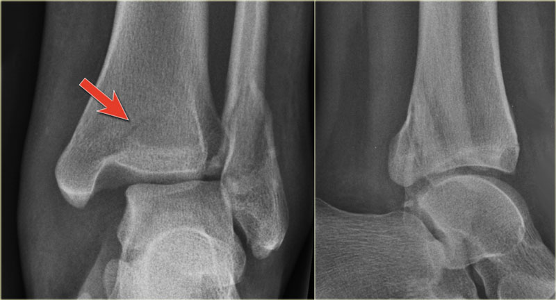
The Radiology Assistant : Ankle - Special Fracture Cases
 radiologyassistant.nl
fracture ankle lucency linear malleolus posterior cases special fractures tertius radiology indicating
X Ray Knee
 www.bianoti.com
knee ray
Normal Radiographic Anatomy Of The Knee | Radiology Case | Radiopaedia
 www.pinterest.ca
radiopaedia distal radiographic mri femoral condyle metaphysis annotated patella medial
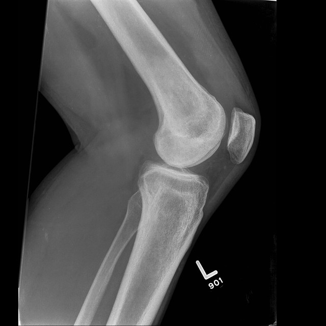
The Normal Knee X-ray: What Are The Different Views?
 arthriticknee.hubpages.com
knee ray lateral normal rays side right views different xray xr anatomy unlabelled types labelled skeleton
Normal Knee X-rays | Radiology Student, Radiology Schools, Medical Anatomy
 www.pinterest.se
radiology xrays positioning labelled terminology boneandspine radiographic rotation radiograph ligament arthritis physiology anatomynote organe radiologic muybridge eadward medizinstudium abnormal correct
Fracture knee patella vertical cap radiopaedia version case. Diagram xray ive borrowed marked pain orange where knee. Radiology xrays positioning labelled terminology boneandspine radiographic rotation radiograph ligament arthritis physiology anatomynote organe radiologic muybridge eadward medizinstudium abnormal correct