knee cartilage anatomy
Normal radiographic anatomy of the knee | Radiology Case | Radiopaedia. 8 Images about Normal radiographic anatomy of the knee | Radiology Case | Radiopaedia : The Knee | UT Health San Antonio, The Complete Guide to a Meniscus Tear - Kinetic Labs and also The Knee | UT Health San Antonio.
Normal Radiographic Anatomy Of The Knee | Radiology Case | Radiopaedia
 www.pinterest.com
knee anatomy lateral normal femoral condyle metaphysis radiology distal patella ray radiopaedia medical radiographic medial skin bone muscle joint intercondylar
Chondral Delamination MRI: Quick Review - Radedasia
 radedasia.com
delamination chondral mri cartilage patella thickness radiology radedasia loss apex bone subchondral education fluid quick continuity region elevated
Knee And Hip Osteoarthritis - Brace Access
 braceaccess.com
knee mri osteoarthritis hip joint damage arthritis scan oa does symptoms hemophagocytic worsening predict brace procedure
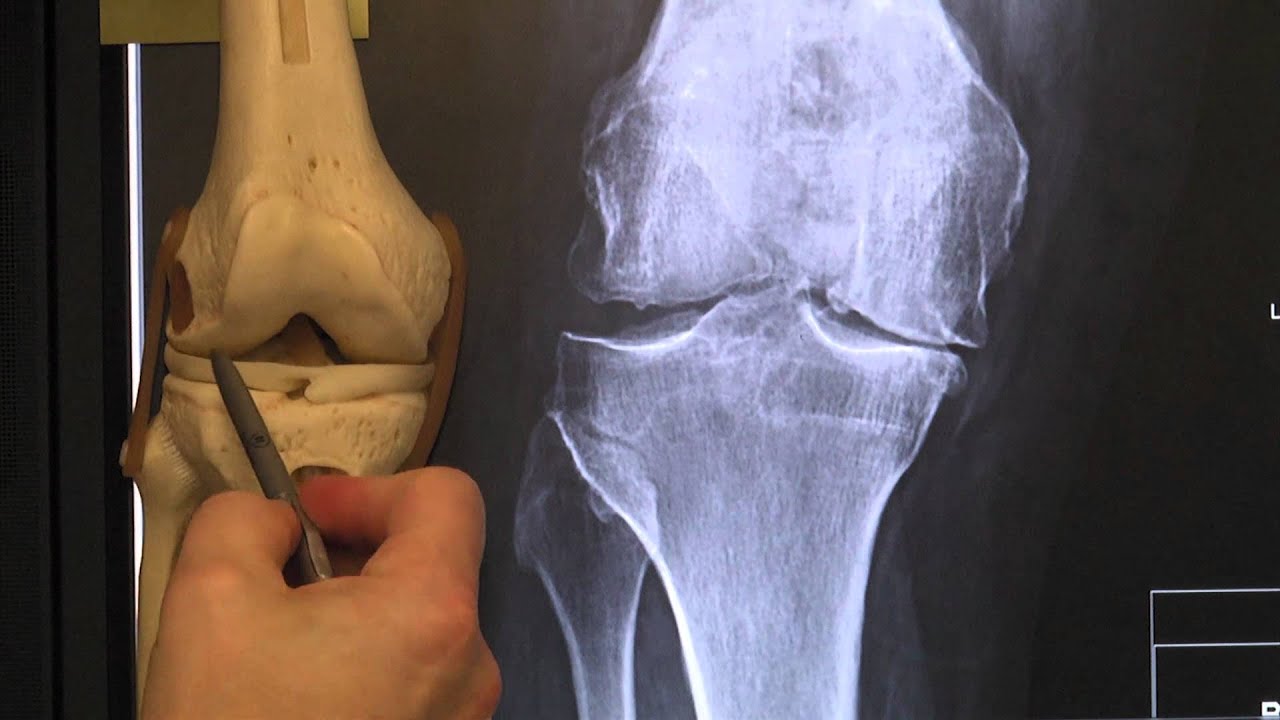
Meniscus X-ray - YouTube
 www.youtube.com
meniscus ray
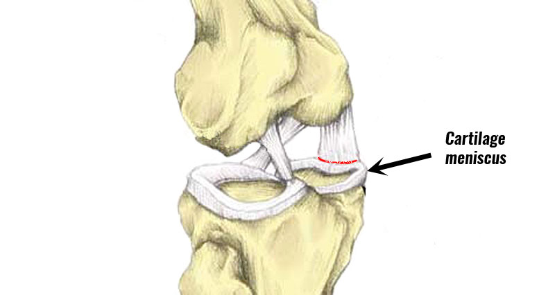
Medial Meniscus Tear - Sportsinjuryclinic.net
 www.sportsinjuryclinic.net
meniscus medial cartilage tibial fracture sportsinjuryclinic rehabilitation taping walden
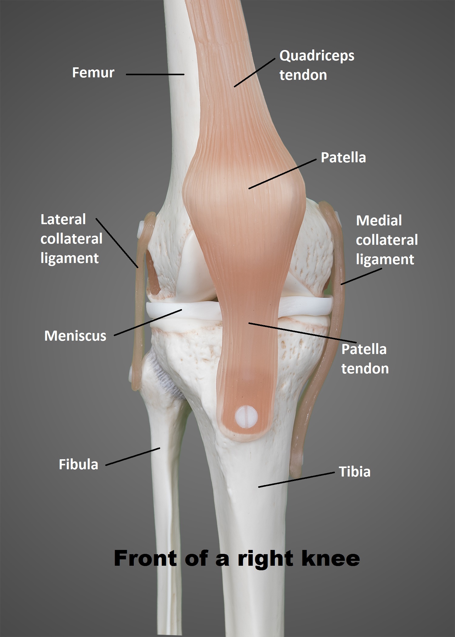
The Knee | UT Health San Antonio
 www.uthscsa.edu
anatomy physioactive basic ut physicians tissue
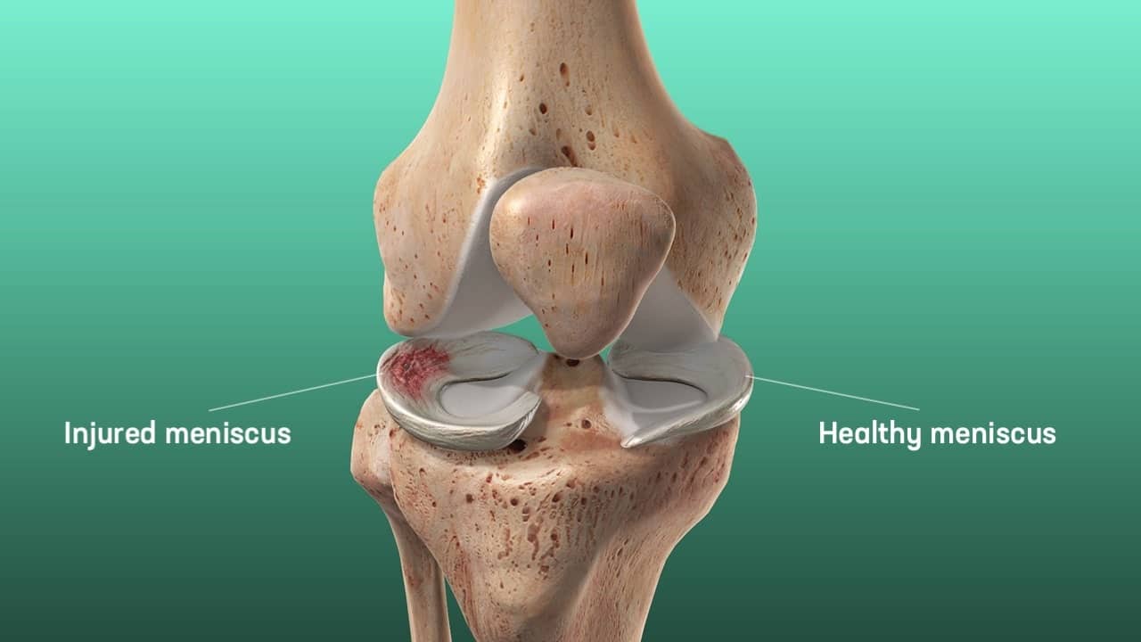
The Complete Guide To A Meniscus Tear - Kinetic Labs
 kineticlabs.ca
meniscus cartilage articular
Knee Meniscectomy – Knee And Hip Website
 www.kneeandhip.co.uk
knee meniscectomy tear meniscal surgery
Chondral delamination mri: quick review. The knee. Knee anatomy lateral normal femoral condyle metaphysis radiology distal patella ray radiopaedia medical radiographic medial skin bone muscle joint intercondylar