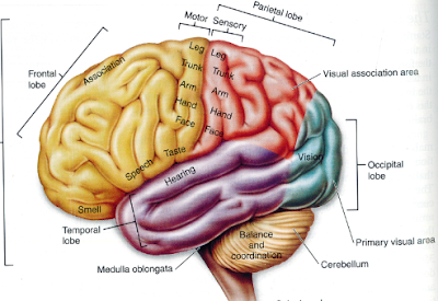
regions of the brain
Patterns of Brain Atrophy That Differentiate Corticobasal Degeneration. 16 Images about Patterns of Brain Atrophy That Differentiate Corticobasal Degeneration : Diagram Brain Regions - Aflam-Neeeak, Medical Illustrations Laura Maaske Medical Illustrator & Biological and also earlier maps.
Patterns Of Brain Atrophy That Differentiate Corticobasal Degeneration
 jamanetwork.com
atrophy corticobasal degeneration supranuclear palsy
Brain Frontal Section 2 - Humpath.com - Human Pathology
 www.humpath.com
brain frontal section sections humpath portfolio human
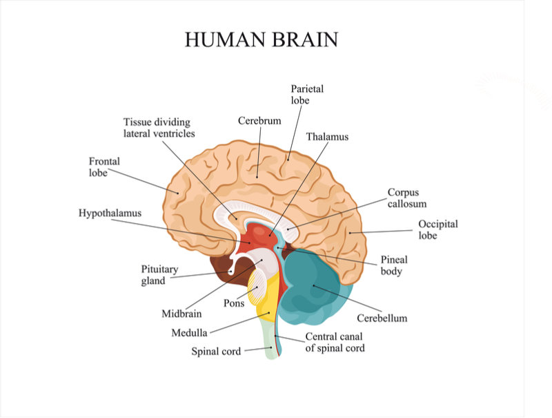
Medical Illustrations Laura Maaske Medical Illustrator & Biological
 medimagery.com
brain regions illustration anatomy medical medimagery illustrations
Neurobiology — Chapter 1: The Brain And Behavior
 biol469.tumblr.com
brain hypothalamus three divided system into regions behavior chapter limbic fornix broader
Earlier Maps
 users.loni.usc.edu
schizophrenia matter loss gray males females
Drinking Alcohol Shrinks Critical Brain Regions In Genetically
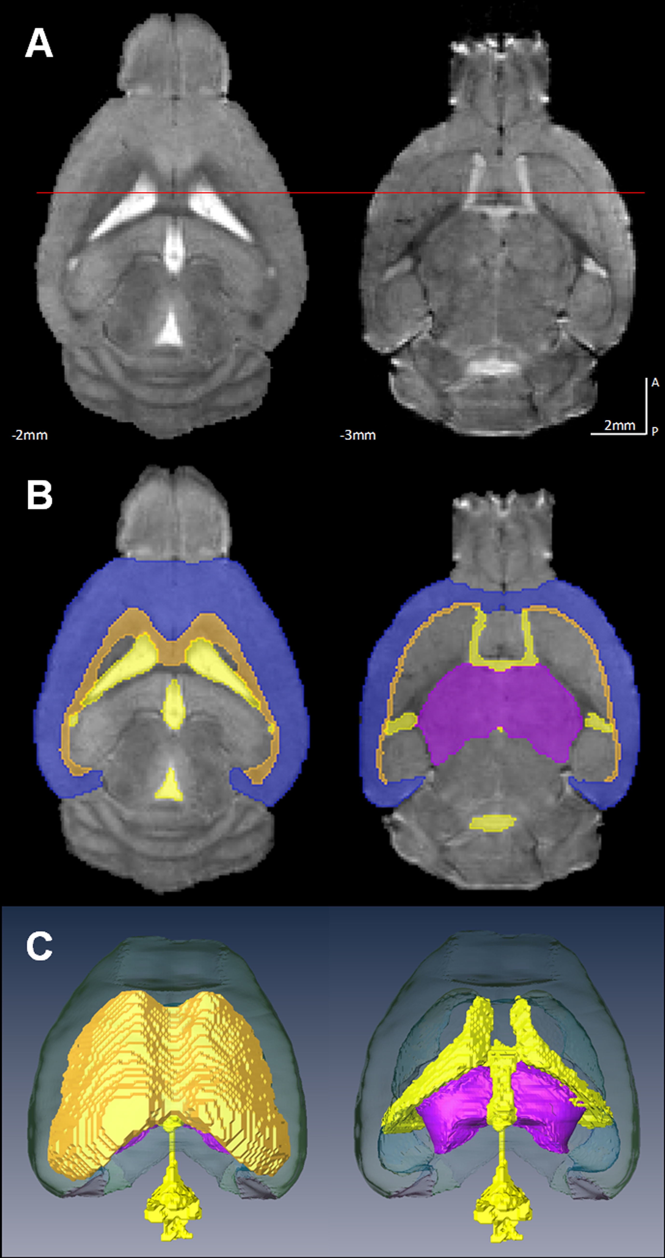 www.bnl.gov
brain mice mouse alcohol regions drinking volume bnl genetics guard against critical genetically vulnerable shrinks february scienceblogs
Clinical And Technical Considerations For Brain PET Imaging For
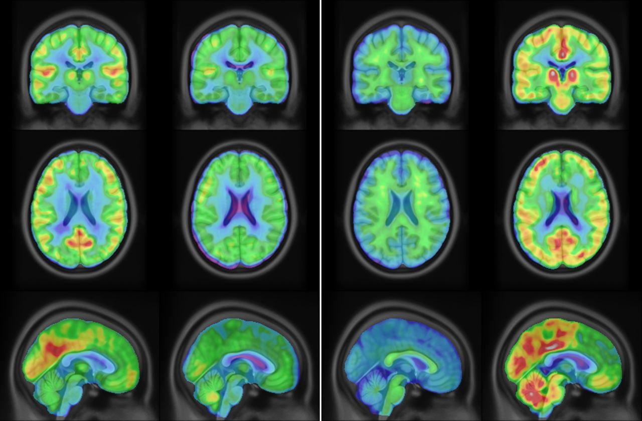 tech.snmjournals.org
considerations fdg snmjournals
Functions Of Brain Regions Quiz
 www.purposegames.com
brain regions functions game
Phone Addiction: Smartphone Use Can Affect Your Brain, Study Says
 www.usatoday.com
phone cell student phones class students distracting cons use smartphone looking cellphones cheat addiction study children teachers texting bring through
Brain Frontal Section 2 - Humpath.com - Human Pathology
 www.humpath.com
brain frontal pathology section humpath
Mathematicians: An Equation Is A Sense Of Beauty | TechieTonics
 www.techietonics.com
mathematicians techietonics mathematical
Diagram Brain Regions - Aflam-Neeeak
 aflam-neeeak.blogspot.com
regions cerebrum stem cerebellum hormone pregnenolone neeeak aflam
What Makes Us Human? Unique Brain Area Linked To Higher Cognitive Powers
 medicalxpress.com
brain human area makes cognitive monkeys frontal similarities cortex areas powers linked unique oxford university higher pars regions opercularis uniquely
Science For All: MAJOR FUNCTIONAL REGIONS OF HUMAN BRAIN
 sciencetable.blogspot.com
regions cortex cerebral
Largest Region Of The Brain Site Of Origin Of Conscious Thought And
 cloudshareinfo.blogspot.com
cloudshareinfo functions
Migraine Patients Have Brain Abnormalities, MRI Scans Show
 www.medicalnewstoday.com
mri migraine abnormalities galvos headache sufferers metus
Earlier maps. What makes us human? unique brain area linked to higher cognitive powers. Schizophrenia matter loss gray males females