wrist anatomy bone
Carpal bones | Radiology Reference Article | Radiopaedia.org. 9 Pictures about Carpal bones | Radiology Reference Article | Radiopaedia.org : TFCC & Ulnar-side wrist injuries — Rayner & Smale, Maxilla: Anatomy, function, clinical aspects | Kenhub and also Surface Anatomy of the Arm Medial View of Caucasian Male | Joel Gordon.
Carpal Bones | Radiology Reference Article | Radiopaedia.org
 radiopaedia.org
carpal radiopaedia radiology
Surface Anatomy Of The Arm Medial View Of Caucasian Male | Joel Gordon
 joelgordon.photoshelter.com
medial
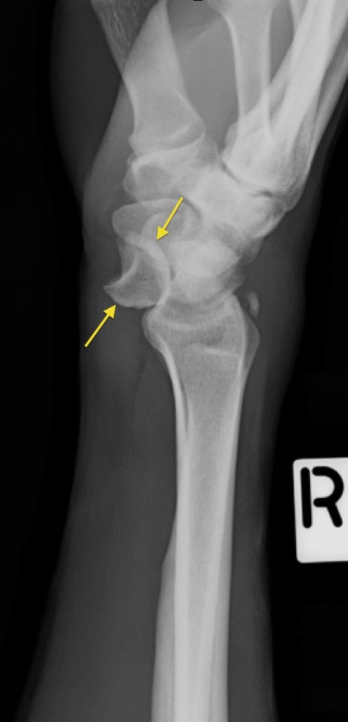
Lunate Dislocation - Radiology At St. Vincent's University Hospital
 www.svuhradiology.ie
lunate dislocation radiology svuhradiology ie
Pin On Radiologic Technologist
 www.pinterest.com
anatomy forearm ray normal xray radiology rad skeletal elbow radiography radiologic tech technologist imaging sickkids
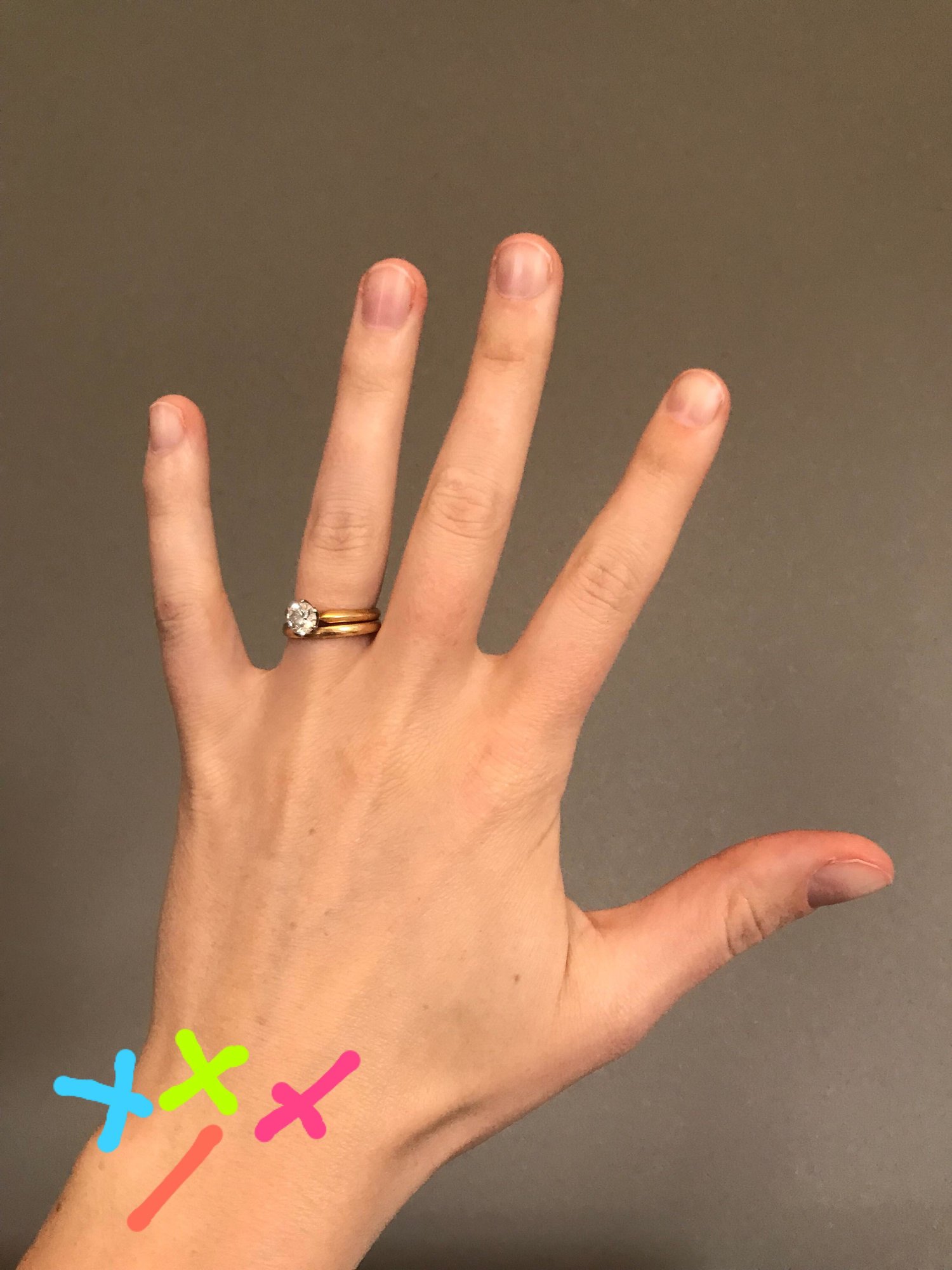
Kendall Marshall’s Broken Wrist – What Is A Scaphoid Bone?
 noelhenley.com
scaphoid fractures kendall sprained sprains torn
Maxilla: Anatomy, Function, Clinical Aspects | Kenhub
maxilla kenhub anatomy function
Kendall Marshall’s Broken Wrist – What Is A Scaphoid Bone?
 noelhenley.com
wrist scaphoid broken ray sprained symptoms vs bone hand fx sprain fractured bones injury marshall kendall where circle yellow around
Bones Of The Head: Skull Anatomy | Kenhub
head bones skull anatomy kenhub
TFCC & Ulnar-side Wrist Injuries — Rayner & Smale
 www.raynersmale.com
wrist ulnar tfcc side druj injuries scapholunate injury supination radial interval pronation
Maxilla kenhub anatomy function. Kendall marshall’s broken wrist – what is a scaphoid bone?. Carpal bones